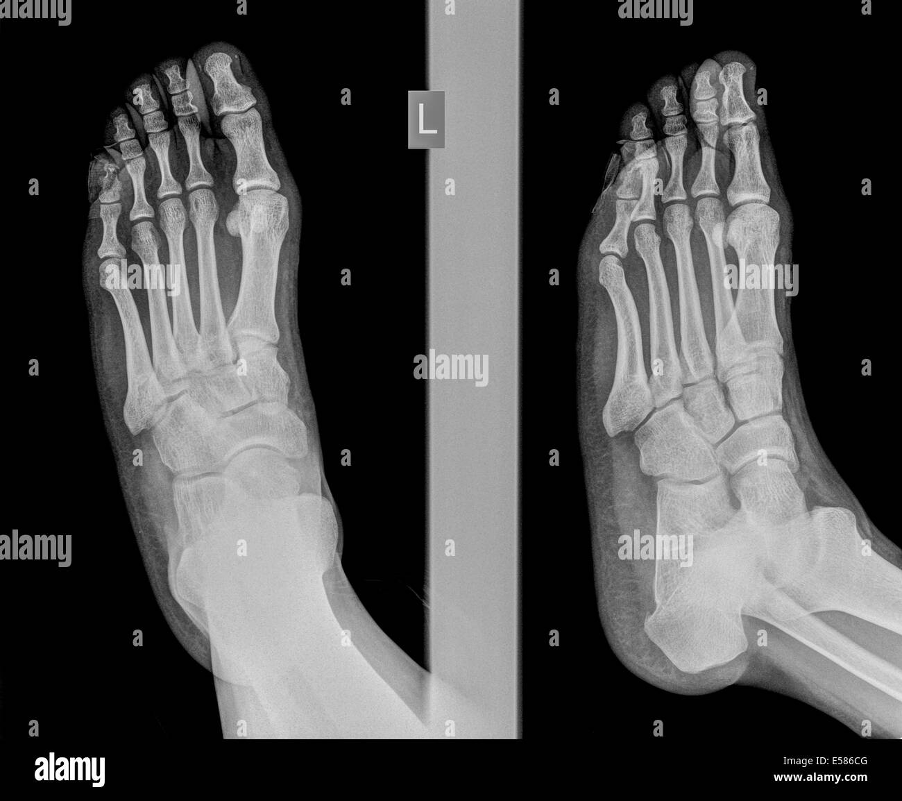
Plain radiograph (AP and lateral oblique) of the left foot (injured
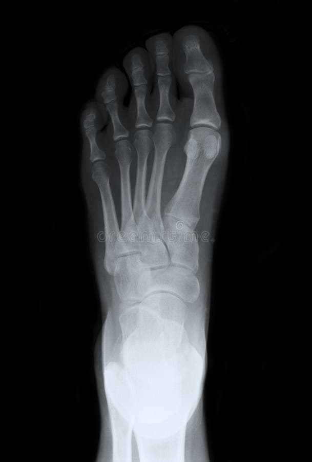
Left Foot Top Xray Royalty Free Stock Image Image 23546186
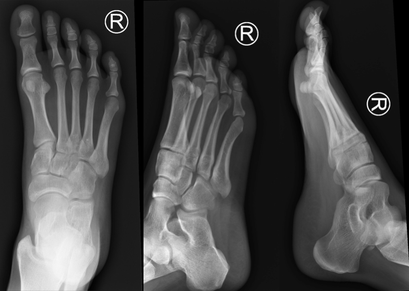
Gender: Female. x-ray. Frontal. Oblique. Lateral. Normal right foot radiographs in a young adult female for reference.

Pin on Xrays
Remember to check the whole film, though. Often, a foot x-ray is also requested for the investigation of osteomyelitis , arthritides , or bone lesion. This article relates mainly to traumatic injuries to the foot. A basic review should start with AP and lateral views (including the entire foot and ankle). With the exception of trauma, these.

www.lisfranc.ca Lisfranc Fracture / Injury Blog Lisfranc / Midfoot
A foot X-ray is a test that produces an image of the anatomy of your foot. Your healthcare provider may use foot X-rays to diagnose and treat health conditions in your foot or feet. Foot X-rays are quick, easy and painless procedures. A radiologic technologist will place your leg on an X-ray table and then take multiple pictures of it.
Episode 76.0 The Lisfranc Injury Core EM
This is a basic article for medical students and other non-radiologists. A foot x-ray, also known as foot series or foot radiograph, is a set of two x-rays of the foot. It is performed to look for evidence of injury (or pathology) affecting the foot, often after trauma.
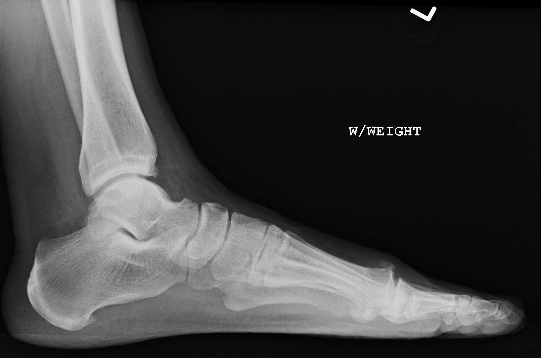
Standing lateral view Xray of the left foot. The os intermetatarseum
What to Expect During Your Foot X-Ray Procedure. Before the x-rays are taken you will be asked to take off your shoes and socks and roll up the legs of your pants. You will need to remove any jewelry or metal objects you may be wearing—for example, an ankle bracelet or toe ring. If you are pregnant, you must let the doctor know before.

Osseous injuries of the foot an imaging review. Part 1 the forefoot
Ankle and foot radiography is the plain radiographic investigation of the distal tibia and fibula, the tarsal bones and metatarsals. Radiographic examination of the foot and ankle are often requested together, however, there is a plethora of literature to aid in the correct request of x-ray examinations in this region including the Ottawa ankle.
21 Treatment of Painful Big Toes Utilizing BioPro HemiImplant
Share this: Chapter 3 Radiology of the Foot and Ankle Orla Doody and Melanie A. Hopper Introduction There are a number of imaging modalities available to the clinician to assist in the evaluation of foot and ankle pathology. An understanding of each technique and its limitations is crucial in providing a rational approach to radiological.

Foot Xray Stock Photo Image 42131008
The bases of the metatarsals and the tarsal bones are the most reliable rotation indicator on the DP view. If the foot is over rotated externally, the metatarsal bases will be heavily superimposed whilst the tuberosity of the navicular bone can be seen in profile. Over rotation internally will open up the metatarsal bases and the resultant.
Normal Left Ankle Xray
26. Hallux sesamoid bones. 27. Lesser metatarsal sesamoid bone (of fifth metatarsal) 28. Second metatarsophalangeal joint. 29. Proximal phalanx third toe. 30.

Left Foot Top Xray Royalty Free Stock Image Image 23546186
"when sitting i favor leaning to my left side, same when working from desk. now after 5 or so years, it's creating lower back discomfort. x-ray clear. i work on couch using laptop, and my way of sitting is left foot on my right new while i am leaning towar" Answered by Dr. Warren Wolfe: Common problem: You have poor body mechanics and your musculature need.
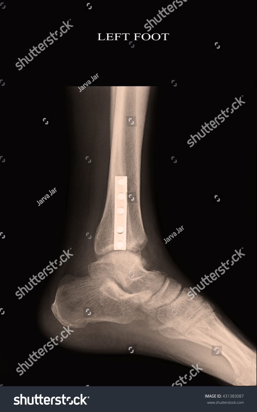
Xray Left Foot Stock Photo 431383087 Shutterstock
Xray left foot show amputaion toe at left foot. Find Left Foot Xray stock images in HD and millions of other royalty-free stock photos, 3D objects, illustrations and vectors in the Shutterstock collection. Thousands of new, high-quality pictures added every day.

Xray left foot metatarsal pain Radiology
Approximately 40 percent of adults in the United States experience foot problems. 1 Plain radiography is an important diagnostic tool in the initial evaluation of patients with chronic foot pain.

normal right foot x ray Google Search Foot x ray
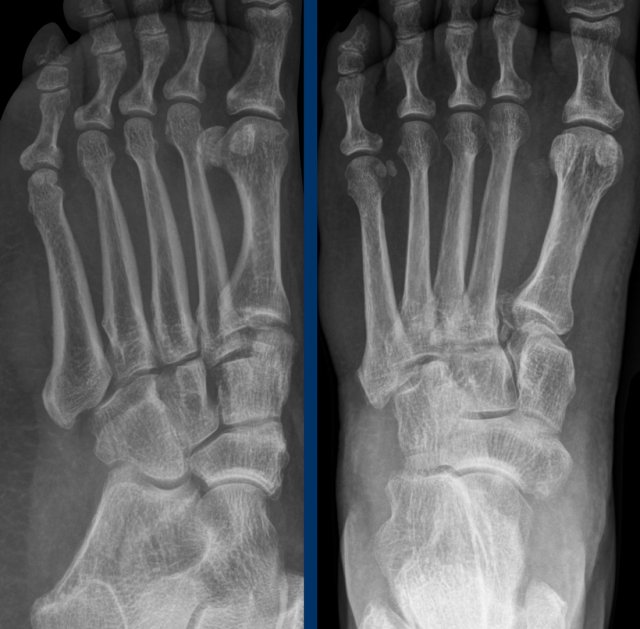
Lisfranc injury. The 'Lisfranc' ligament stabilises the mid-forefoot junction. Loss of alignment of the 2nd metatarsal base with the intermediate cuneiform indicates injury to this important ligament. Every post-traumatic foot X-ray must be checked for loss of alignment at the midfoot-forefoot junction (tarsometatarsal joints).
/x-ray-image-of-bone-fracture-at-5th-metatarsal-left-foot-945203958-140a7bb8add94610838f0b3632543a5c.jpg)
Jones Fracture of the Foot Symptoms, Treatment, and Recovery
No fracture is seen. Bones show normal alignment and architecture. Joint spaces and articular margins are intact. Soft tissues show normal appearance. FREE download PDF Word format X rays Left Foot AP/OBL . Also available other updated Radiology MRI, CT Scan, Xray, Sonography, USG, Mammography, PET CT, EEG and ECG Report templates.
xray of a foot showing a fracture in the intermediate phalanx of the
Download scientific diagram | Left foot X-ray: (a) Anteroposterior view; (b) lateral view; (c) oblique view and (d) axial calcaneus view. Note the gross talar head irregularity with dense areas.

Xray Left Foot Stock Photo 434091277 Shutterstock
A foot X-ray is a test that produces an image of the anatomy of your foot. Your healthcare provider may use foot X-rays to diagnose and treat health conditions in your foot or feet. Foot X-rays are a simple, quick, and painless process. Your leg will be positioned on an X-ray table by a radiologic technician who will then take numerous images.
