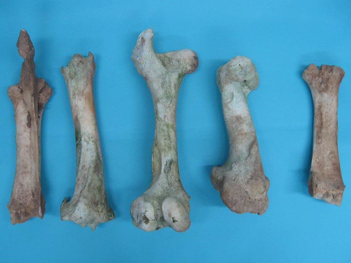
Cow Bones, Loose Cow Bones

Complete Cow Hind leg with Pathology (Bos taurus) Cow, Pathology, Taurus
In the cow the tuber coxae is visible and is readily palpable. The sacral tuber has two prominences; the cranial and caudal dorsal iliac spines. The iliac crest is thin and concave. The ileal wing is orientated in a vertical manner. Ischium The ischial tuberosity is triangular in shape. Femur
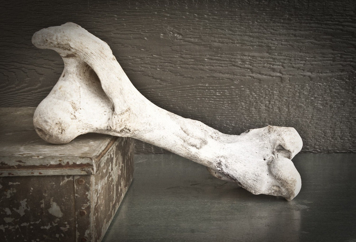
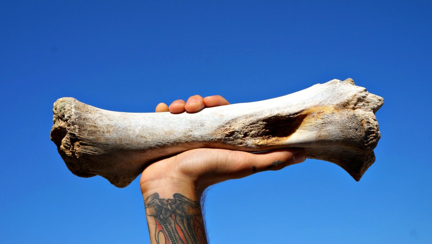
Bleached Cow Leg Bone
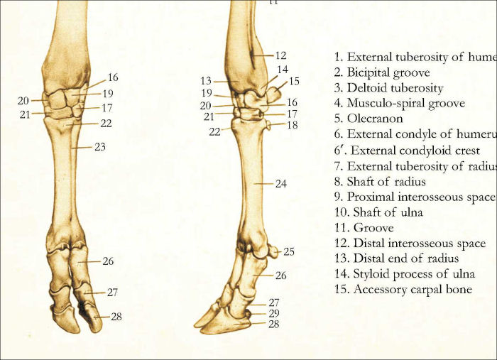
Bovine Limb Anatomy. Home. 3D. Radiographic Projection. Tarsus (Left) Lateromedial (Juvenile) Dorsoplantar (Juvenile) Lateromedial (Mature) Dorsoplantar (Mature)

Cow legs Horse anatomy, Large animal vet, Animal paintings
A side of beef is literally one side of the beef carcass that is split through the backbone. Each side is then halved between the 12th and 13th ribs. These sections are called the forequarter (front of the cow) and the hindquarter (back of the cow).

Set of 8 Cow Leg Bones , Real Animal Bones , Gothic Pagan Decor , Cow
In the front of the cow, from the front legs to the head, a diagram of the cow's skeletal system includes the cannon, knee joint, radius, sternum, elbow joint, ulna, humerus, shoulder joint, shoulder blade and eye socket. From the top of the head and along the top side of the cow, the skeletal system includes the horn cones, cervical.

Cow Leg Bones Diagram Vintage Cow and Bull Hind Quarters View
25/04/2023 28/10/2022 by Sonnet Poddar The cow leg anatomy consists of bones, muscles, nerves, and vessels. Bones are the hardest and main component of the cow leg structure. Again, the muscles are also essential as most vessels and nerves pass along or within them.

Cow Bones, Loose Cow Bones
Table of Contents Cow anatomy External body parts of a cow Little on terminologies of cow body Internal body parts of a cow Cow anatomy bones Bones of thoracic limb of a cow Bones from the hindlimb of a cow Cow skull anatomy Vertebrae of a cow Ribs and sternum anatomy of a cow Cow muscle anatomy Muscles of thoracic limb of a cow
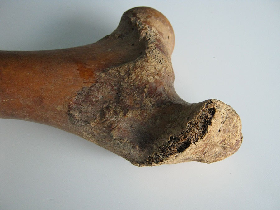
Weathered Cow Leg Bone by cellarandshed on Etsy
1 Overview 2 Bones 2.1 Metacarpals and Metatarsals 2.2 Phalanges 3 Joints 3.1 Metacarpophalangeal/Fetlock Joint 3.2 Proximal Interphalangeal/Pastern Joint 3.3 Distal Interphalangeal/Coffin Joint 4 Musculature 4.1 Forelimb 4.2 Hindlimb 5 Vasculature 6 Innervation 7 The Bovine Claw 8 Webinars Overview Cattle are artiodactyl unguligrade animals.

Cow Leg Bones Diagram / 6.4 The Forelimb Medicine LibreTexts Bones
3. The Omasum - this part of the stomach is a 'filter'. It filters through all the food the cow eats. The cud is also pressed and broken down further. 4. The Abomasum - this part of the stomach is like a humans stomach and is connected to the intestines. Here, the food is finally digested by the cows stomach juices and essential.
Skeletal Anatomy of the Cow Poster
There are generally three levels of identification that can be utilized to distinguish between human and non-human animal bones: 1) gross skeletal anatomy, 2) bone macrostructure, and 3) bone microstructure (histology).

Identification cattle hock bone
Cow Shoulder Blade: R-1172-20: Cow Leg Bones: R-1172-40: Cow Vertebra: R-1172-41: Connected Cow Vertebrae: R-1172-50: Cow Pelvic Bone : R-1172-LB: Assorted Cow Bones: Genus and species: Bos taurus. Ranch. Cows are not an endangered species and are not subject to CITES controls.

Femur Bone Of Cow Isolated On A White Background Stock Image Image of
Bovine Lameness and Podiatry Bovine Foot Anatomy Bones and joints While the cannon bone of a horse is MCIII or MTIII, in a cow it is a fused MCIII+IV or MTIII+IV. The fusion is present at the fetlock joint and above. Cattle do have the same bones and joints as horses below the fetlock but in duplicate form.

Cow Leg Bones Diagram / Human Skeleton Labeled Diagram . Human Skeleton
This veterinary anatomical atlas includes 27 scientific illustrations with a selection of labelled structures to understand and discover animal anatomy (skeleton, bones, muscles, joints and viscera). Positional and directional terms are also illustrated.
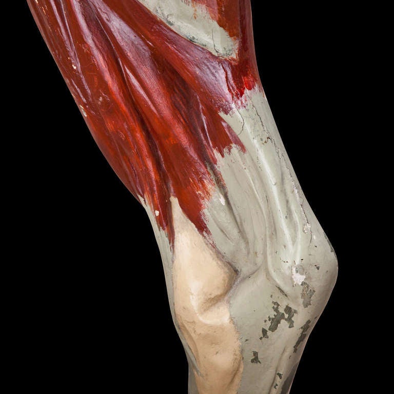
Anatomical Model of Cow Leg at 1stdibs
It is a synovial type of joint responsible for extension and flexion movements of the cattle's hind limb. I will describe the bony involvement and articulations of the cow's hock with their binding materials (ligaments). You will also find a short description of the muscles from this guide that are forming cattle's hock.
Cow Leg Bone
The forelimbs of a cow consist of the humerus, radius, ulna, carpal bones, metacarpal bones, and phalanges. The humerus is the long bone situated between the scapula and the elbow joint. The radius and ulna are the bones that run from the elbow joint to the carpus or knee joint.
Cow Leg Bones Diagram / Anterior view of left tarsal bone and ankle
Typically, fractures result from external trauma to the limb. Causes of the fracture vary depending on age. Even before delivery, calves are subject to fractures due to dystocia (backward presentation, or an oversized calf). Newborn calves may suffer from being stepped on by a cow, and as they grow larger, fractures can occur from either self.

il_fullxfull.220623459.jpg (726×613) Animal Bison Pinterest
Cattle muscles anatomy How many muscles does a cow have? Identification of forelimb muscles from the cow Muscles of the arm region of a cow Muscles of the forearm of the cow Muscles of the hindlimb of a cow Lateral muscles of the hip and thigh of a cow Cranial muscle of the cow's thigh Medial muscles of the cow's thigh Cow leg muscle anatomy
