Anatomy of the Foot and Ankle OrthoPaedia
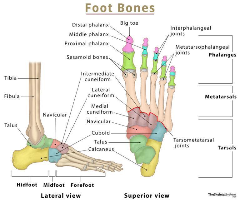
Foot Bones Names, Anatomy, Structure, & Labeled Diagrams
The foot itself can be divided into three sections: the hindfoot, midfoot and forefoot and the foot bones can be grouped into three sets: the tarsal bones, the metatarsals and the phalanges .

Foot Bone Anatomy Vector Illustration 539973 Vector Art at Vecteezy
It consists of 28 bones, which can be divided functionally into three groups, referred to as the tarsus, metatarsus and phalanges. The foot is not only complicated in terms of the number and structure of bones, but also in terms of its joints.

Ankle and Foot Pain Massage Therapy Connections
1/3 Synonyms: Ossa metatarsalia The metatarsal bones are a group of five long bones located in the metatarsus of the foot, between the tarsal bones (near the ankle) and the phalanges (toe bones).
Structure of skeleton of the foot, Tarsals, Metatarsals and Phalanges Science online
The diagram of bones in the ankle and foot is given below: Tarsal Bones The tarsal bones in the foot are located amongst tibia, metatarsal bones, and fibula. There are in all 7 bones, which fall under tarsal bones category. They are: Calcaneus or Calcaneum: To explain the term in layman's language, it is the heel bone in the skeletal system.

The bones in the foot inferior view (Picture illustrated from Thieme... Download Scientific
The foot is a complex structure comprised of over 26 bones, 30 joints, numerous tendons, ligaments, and muscles responsible for our ability to stand upright, supporting the weight of the entire body and provides the base for the mechanism for bipedal gait. The foot corresponds to the portion of the lower extremity distal to the ankle and divides into hind, mid and forefoot.
.jpg)
Foot Bone Diagram resource Imageshare
The foot structure is complex, consisting of many bones, joints, ligaments and muscles. The foot is divided into three parts: rearfoot, midfoot, and forefoot. A clinician's ability to understand the anatomical structures of the foot is crucial for assessment and treatment, especially for clinicians working with clients with musculoskeletal conditions.
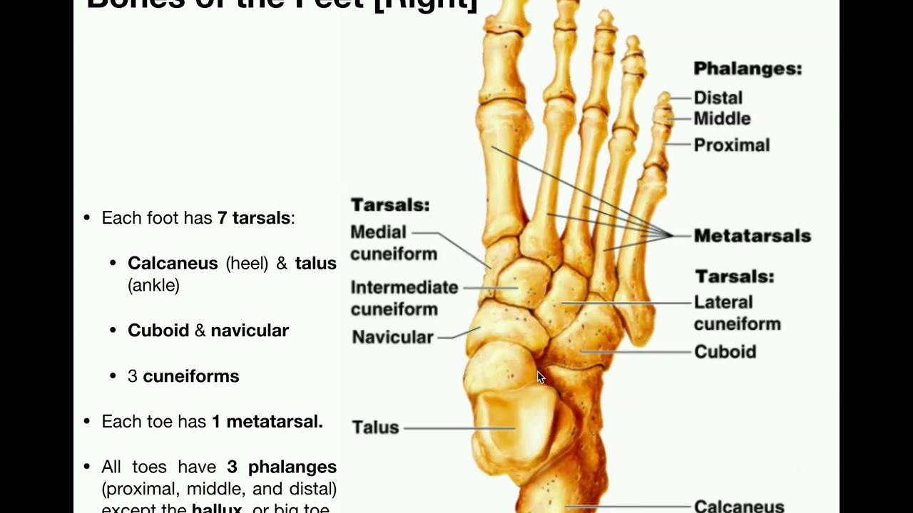
Anatomy Specific Bones of the Feet YouTube
Foot Anatomy The foot contains 26 bones, 33 joints, and over 100 tendons, muscles, and ligaments. This may sound like overkill for a flat structure that supports your weight, but you may not realize how much work your foot does!
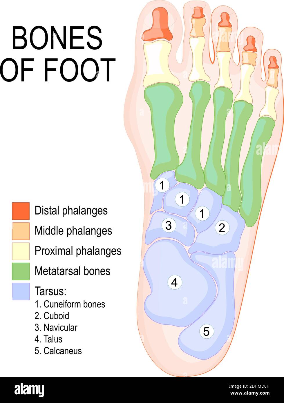
Bones of foot. Human Anatomy. The diagram shows the placement and names of all bones of foot
Human body Foot Foot The foot is the lowermost point of the human leg. The foot's shape, along with the body's natural balance-keeping systems, make humans capable of not only walking, but.

Anatomy of the Foot and Ankle OrthoPaedia
Figure 1: Bones of the Foot and Ankle Regions of the Foot The foot is traditionally divided into three regions: the hindfoot, the midfoot, and the forefoot (Figure 2). Additionally, the lower leg often refers to the area between the knee and the ankle and this area is critical to the functioning of the foot.

anatomy of the foot Ballet News Straight from the stage bringing you ballet insights
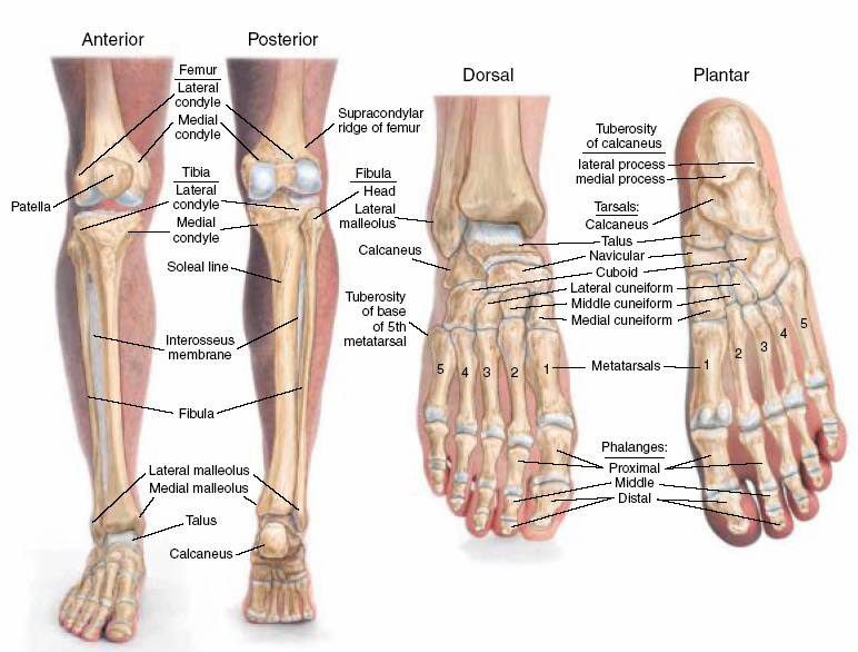
The talus is on top of the foot and forms a joint with the tibia and fibula, of the lower leg. The calcaneus is underneath the talus and forms the heel bone. There is also one intermediate.

Foot & Ankle Bones
Summary The foot is an intricate part of the body, consisting of 26 bones, 33 joints, 107 ligaments, and 19 muscles. Scientists group the bones of the foot into the phalanges, tarsal.

Foot Description, Drawings, Bones, & Facts Britannica
Foot Bones: Forefoot. The forefoot consists of 19 bones; 5 metatarsal bones and 14 phalanges. The big toe has 2 phalanges bones, while the remaining four have 3 phalanges each. The 1st metatarsal is the shortest and thickest of the metatarsals, and it is designed to take up to 40% of your body weight in standing, which rises to 70% when walking.

Foot bones anatomy Royalty Free Vector Image VectorStock
The first metatarsal bone leads to the big toe and plays an important role in forward movement. The second, third, and fourth metatarsal bones provide stability to the forefoot. Sesamoid bones: These are two small, oval-shaped bones beneath the first metatarsal on the underside (plantar surface) of the foot. It is embedded in a tendon at the.

Diagram showing metatarsal bones of the foot. Download Scientific Diagram
The bones of the foot provide mechanical support for the soft tissues; helping the foot withstand the weight of the body whilst standing and in motion. They can be divided into three groups: Tarsals - a set of seven irregularly shaped bones. They are situated proximally in the foot in the ankle area. Metatarsals - connect the phalanges to.
Anatomy The Bones Of The Foot
Human body Skeletal System Bones of foot Bones of foot The 26 bones of the foot consist of eight distinct types, including the tarsals, metatarsals, phalanges, cuneiforms, talus,.

Foot skeleton composed of 28 skeletal bones. It can be divided into... Download Scientific Diagram
The anatomy of the foot The foot contains a lot of moving parts - 26 bones, 33 joints and over 100 ligaments. The foot is divided into three sections - the forefoot, the midfoot and the hindfoot. The forefoot This consists of five long bones (metatarsal bones) and five shorter bones that form the base of the toes (phalanges).
