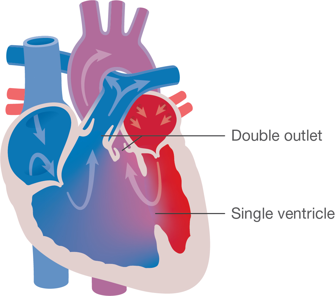
Double Outlet Right Ventricle (DORV) Little Hearts Matter

Surgically Constructed DoubleOutlet Right Ventricle Circulation
A double-chambered right ventricle (DCRV) is a heart defect, typically congenital, in which the right ventricle (RV) is separated into a proximal high-pressure (anatomically lower) chamber and distal low-pressure (anatomically higher) chamber [1].
Double Outlet Right Ventricle (DORV) Little Hearts Matter
Double-chambered right ventricle (DCRV) is an uncommon congenital malformation in which anomalous muscle bundles dissect the RV into two chambers. It is commonly associated with other congenital anomalies, most frequently perimembranous ventricular septal defect (PM-VSD).

Bands in the Heart Multimodality Imaging Review RadioGraphics
Updated: Nov 15, 2022 Author: Shubhayan Sanatani, MD, FRCPC, FHRS; Chief Editor: Stuart Berger, MD more. Approach Considerations Symptoms of double-chambered right ventricle (DCRV) that.

Kiba DoubleChambered Right Ventricle Mount Pleasant Vet Group
Double-chambered right ventricle (DCRV) was first described in 1858 by TB Peacock, but it is now understood to be a form of congenital heart disease wherein there is a mid-cavitary obstruction that divides the right ventricle into a high-pressure proximal portion and a low-pressure distal portion.

Postdelivery graphic anatomy DORV OB Images
Double-chambered right ventricle is a rare congenital or acquired cardiac abnormality and may be associated with other malformations including membranous ventricular septal defect or double outlet right ventricle. 1 Patients may present with symptoms resembling ischemia or heart failure, including dyspnea and acute drops in blood pressure with s.
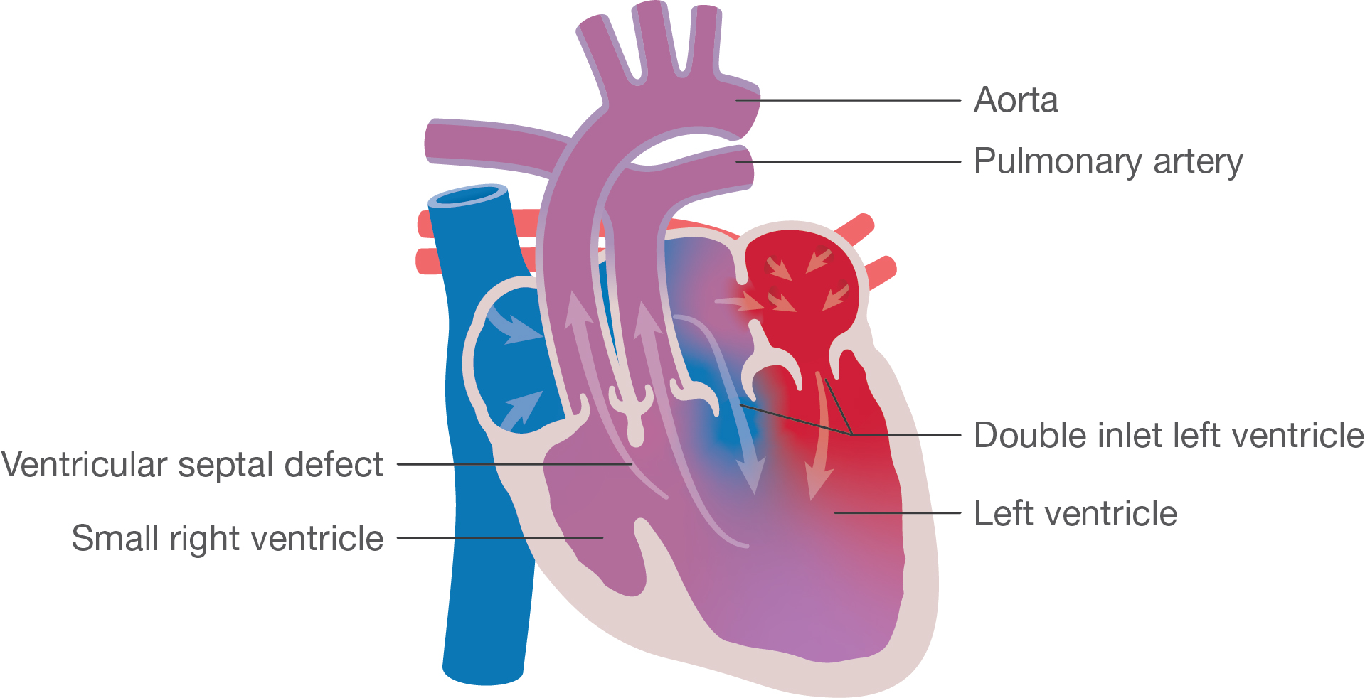
Double Inlet Left Ventricle (DILV) Little Hearts Matter
Double-chambered right ventricle is a congenital anomaly in which the right ventricle is divided into 2 portions by anomalous muscle bundles. These cases often present in children, but rarely in adults. We discuss 2 cases of double-chambered right ventricle, in patients aged 42 and 35 years.
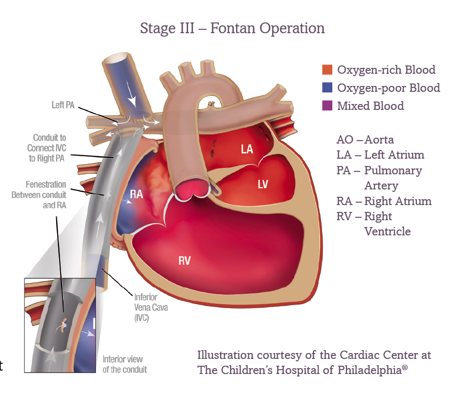
Single Ventricle Defects and the Fontan ACHA
Double-chambered right ventricle is a rare congenital disease frequently misdiagnosed in the adult patient. An anomalous muscle band divides the right ventricle in two cavities causing variable degree of obstruction. Although echocardiography is considered a useful method for the diagnosis of this pathology in children, it has been recognized the transthoracic scanning limitation in adults.

Assessment of Double Chamber Right Ventricle by Resonance Imaging Circulation
Double-chambered right ventricle is a rare congenital heart disorder involving 2 different RV pressure compartments that is often associated with malalignment VSD. Usually, the obstruction is caused by an anomalous muscle bundle crossing the RV from the interventricular septum to the RV free wall.
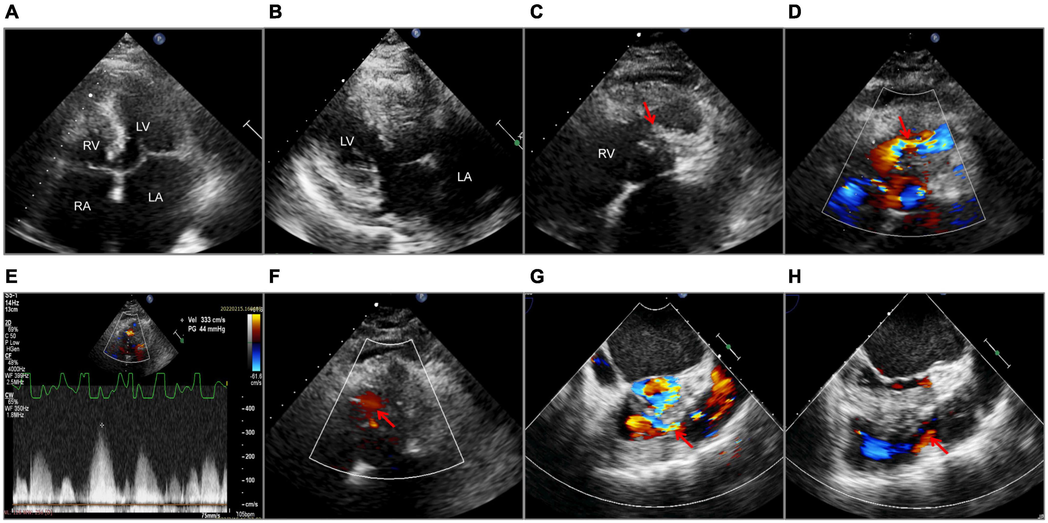
Frontiers Case report Doublechambered right ventricle diagnosed in a middleaged female with
Figure 2. Double-chambered right ventricle. VSD indicates ventricular septal defect; RV, right ventricle; LV, left ventricle. This is an interesting case because these patients usually present with symptoms in infancy or adolescence secondary to right ventricular failure due to the gradient between the 2 RV chambers or associated lesions (ventricular septal defect or subaortic stenosis).

DoubleChambered Right Ventricle and Situs Inversus With Dextrocardia Circulation
Double-chambered right ventricle (DCRV) occurs in approximately 1% of patients with congenital heart disease. The right ventricle (RV) is divided by anomalous muscle bundles into a higher-pressure proximal chamber and lower-pressure distal chamber. The physiology is defined by right ventricular pressure overload of the RV inflow chamber.
Double Outlet Right Ventricle Youth Zone Little Hearts Matter
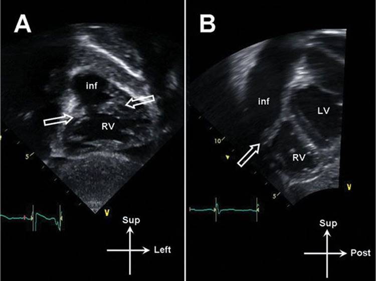
Double-Chambered Right Ventricle. Subcostal right anterior oblique (RAO) echocardiograph view with color Doppler demonstrating ventricular septal defect jet to proximal chamber. (*) =.

Double chambered right ventricle with severe calcification of the tricuspid valve in an elderly
Double-chambered right ventricle is a rare congenital heart disorder involving 2 different RV pressure compartments that is often associated with malalignment VSD. Usually, the obstruction is caused by an anomalous muscle bundle crossing the RV from the interventricular septum to the RV free wall.
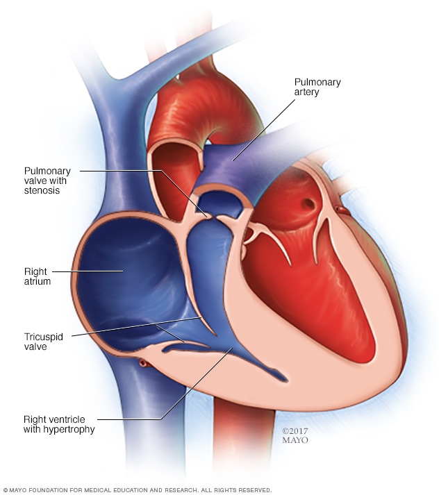
Pulmonary valve stenosis Symptoms and causes Mayo Clinic
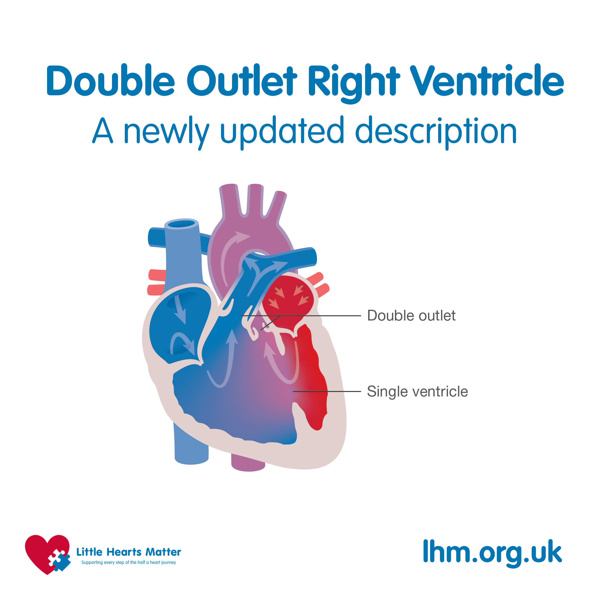
Double-outlet right ventricle is a heart condition present at birth. That means it's a congenital heart defect. In this condition, the body's main artery and the lung artery do not connect to the usual areas in the heart. The body's main artery is called the aorta. The lung artery is called the pulmonary artery.
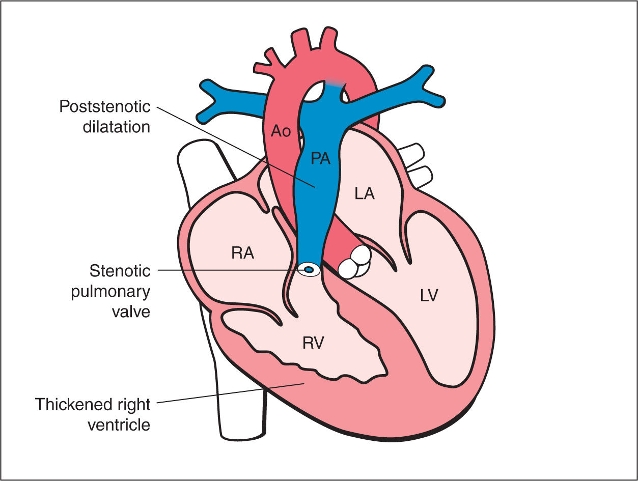
Pulmonary Stenosis, Pulmonary Atresia with Intact Ventricular Septum, and Ductus Arteriosus
A double-chambered right ventricle is a rare heart defect in which the right ventricle is separated into a high-pressure proximal and low-pressure distal chamber. This defect is considered to be congenital and typically presents in infancy or childhood but has been reported to present rarely in adults.
Abnormalities of Right Ventricular Outflow Echocardiography in Pediatric and Adult Congenital
Double-chambered right ventricle is better understood as a form of septated right ventricle (RV) caused by the presence of abnormally located or hypertrophied muscular bands. The.
Frontiers Case report Doublechambered right ventricle diagnosed in a middleaged female with
Double outlet right ventricle (DORV) describes a heart with two major arteries linking to its right ventricle (heart chamber). Normally, only one of these arteries connects to each ventricle. The double link is a rare, congenital (since birth) heart issue. Surgery repairs the problem, but children born with DORV need lifelong follow-up care.
