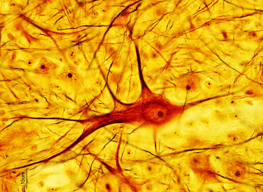
Histology, microscopy, anatomy and disease Week 4 Figure 6 Neurons visualised with a

GolgiCox staining for adult mouse brain. Neurons in all brain... Download Scientific Diagram
Silver impregnation identification of Golgi bodies was discovered in owl optic nerve. Staining reagents since the late 1800s were widely used across all disciplines and for nerve tissue and became a key contributor to advancement in nerve-related research.
Santiago Ramón y Cajal and Camillo Golgi Inside Science Visionlearning
During the years, Golgi technique has undergone many modifications, enhancements and refinements, in order to attain the maximal visualization of neurons and neuronal processes, the minimal precipitations and the reasonable abbreviation of the total time that required for the procedure (Zhang et al., 2003).
Structure of a Neuron Owlcation
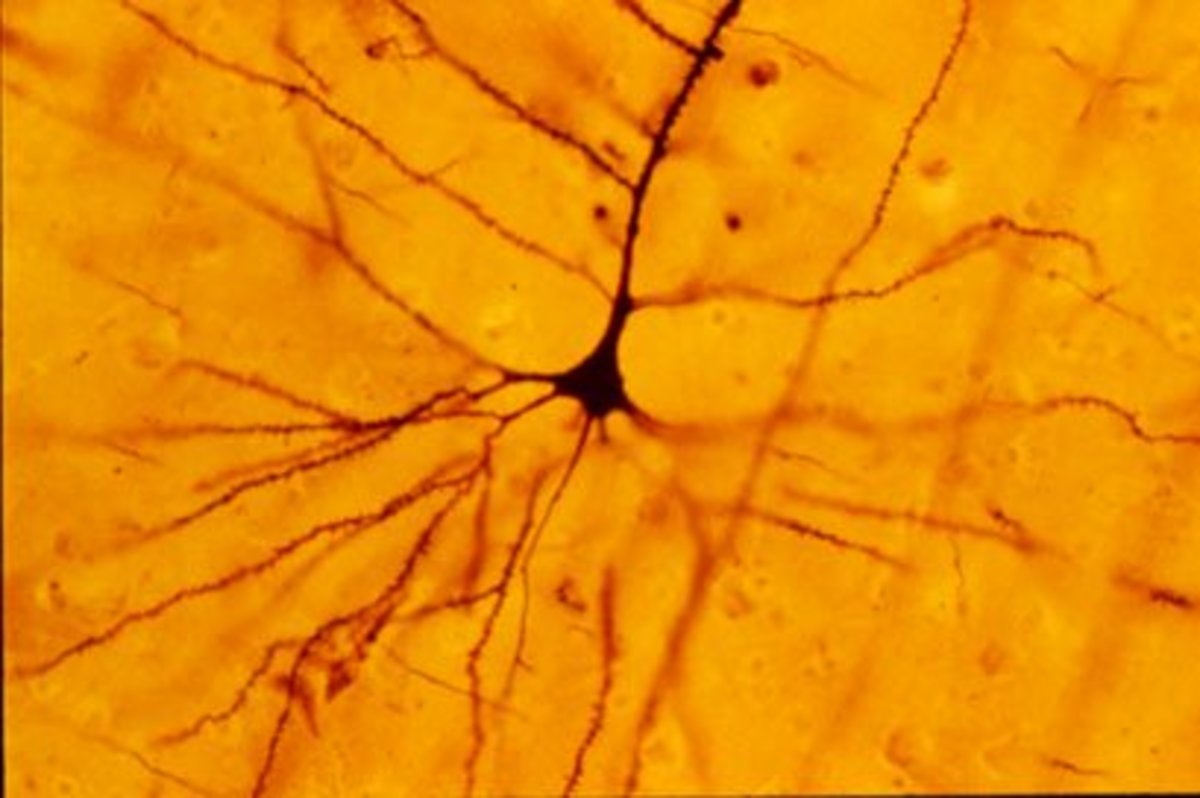
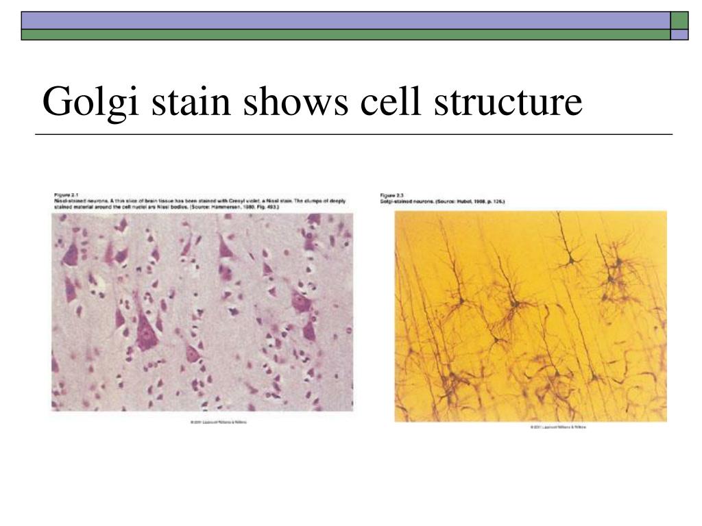
Golgi used his stain to characterize the structure of different types of neurons and discovered a new cell organelle, the Golgi Apparatus. Scientists still use the Golgi stain today, more than 170 years after its advent. Under the microscope, scientists can see details as fine as dendritic spines on Golgi-stained neurons. About the Author

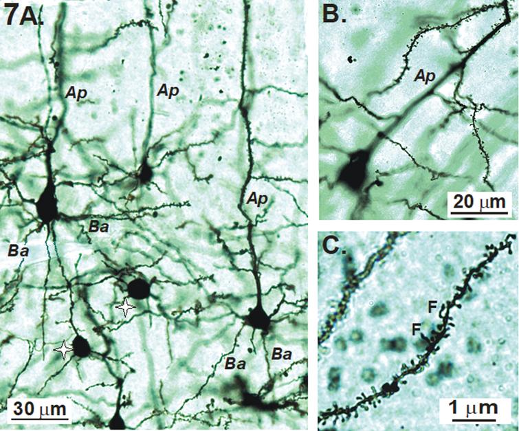
Photomicrographs of Golgistained mouse cortical neurons from slices.... Download Scientific
Golgi Staining A major advancement in the study of neuronal morphology came about in the late 1800s.

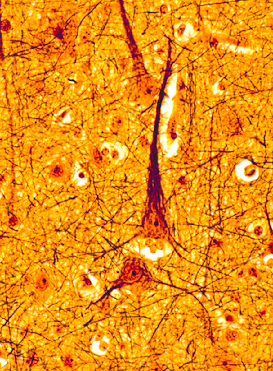
Photomicrographs of Golgistained neurons in primary motor (AC,F,G)... Download Scientific
Golgi staining was first discovered by Camillo Golgi in 1873, called the "black reaction," and to this day remains a reliable method to assess neuronal cytoarchitecture.. (PSD95) for postsynaptic terminals (or dendritic spines). These do not, however, offer the unique advantage of Golgi in staining the entire neuron structure including.

Morphology of Golgistained mediumsized spiny striatal neurons. A... Download Scientific Diagram
The Golgi-Cox method has been one of the most effective techniques for studying the morphology of neuronal dendrites and dendritic spines. However, the reliability and time-consuming process of Golgi-Cox staining have been major obstacles to the widespread application of this technique. To overcome.
Morphometric Analysis of Hippocampal and Neocortical Pyramidal Neurons in a Mouse Model of Late
The Golgi staining technique, also called the black reaction after the stain's color, was developed in the 1870s and 1880s in Italy to make brain cells (neurons) visible under the microscope. Camillo Golgi developed the technique while working with nervous tissue, which required Golgi to examine cell structure under the microscope. Golgi improved upon existing methods of staining, enabling.

A. Golgi cox stained dendrites of CA1 neurons of hippocampus in CCH... Download Scientific Diagram
The neuron doctrine was based on two contributions; Golgi's stain and Cajal's histological studies. The neuron doctrine was named and popularized by Heinrich Wilhelm Gottfried von Waldeyer-Hartz [ 3 ], who coined the name neuron to refer to the nerve cell. The early background: nerve fibres and nerve cells
Histology, microscopy, anatomy and disease Week 4 Figure 6 Neurons visualised with a
Golgi staining allows for the analysis of neuronal arborisations and connections and is considered a powerful tool in basic and clinical neuroscience. The fundamental rules for improving neuronal staining using the method are not fully understood; both intrinsic and extrinsic factors may control the staining process.
PPT Neurons and Glia PowerPoint Presentation ID303331
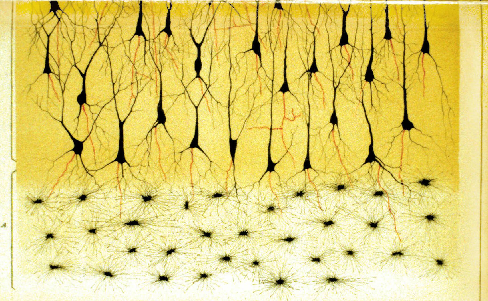
The Golgi staining method was used by the Spanish scientist Ramon y Cajal when he proposed the idea that neurons are not all physically connected, but rather distinct, separate units. This concept is called the neuron doctrine. The major advantage of the Golgi stain is its ability to completely fill out the morphology of the cell.

Golgi stain to see neurons Neuroscience art, Brain art, Art
The Golgi stain has an important place in the history of neuroscience. In the late nineteenth and early twentieth centuries, Santiago Ramón y Cajal used this technique for his seminal studies of the cellular morphology of the nervous system.

GolgiCox staining of different type neurons in the different regions... Download Scientific
Highlights We describe a simple modification of the Golgi-Cox method to differentially stain neurons and glia. If the rat brain or brain tissue block was exposed to a fixative at any stage, glial cells were stained, whereas in its absence neurons were stained. Same brain (different portions) may be used for differential staining of neurons and glia. Staining was achieved in less than 48 h.
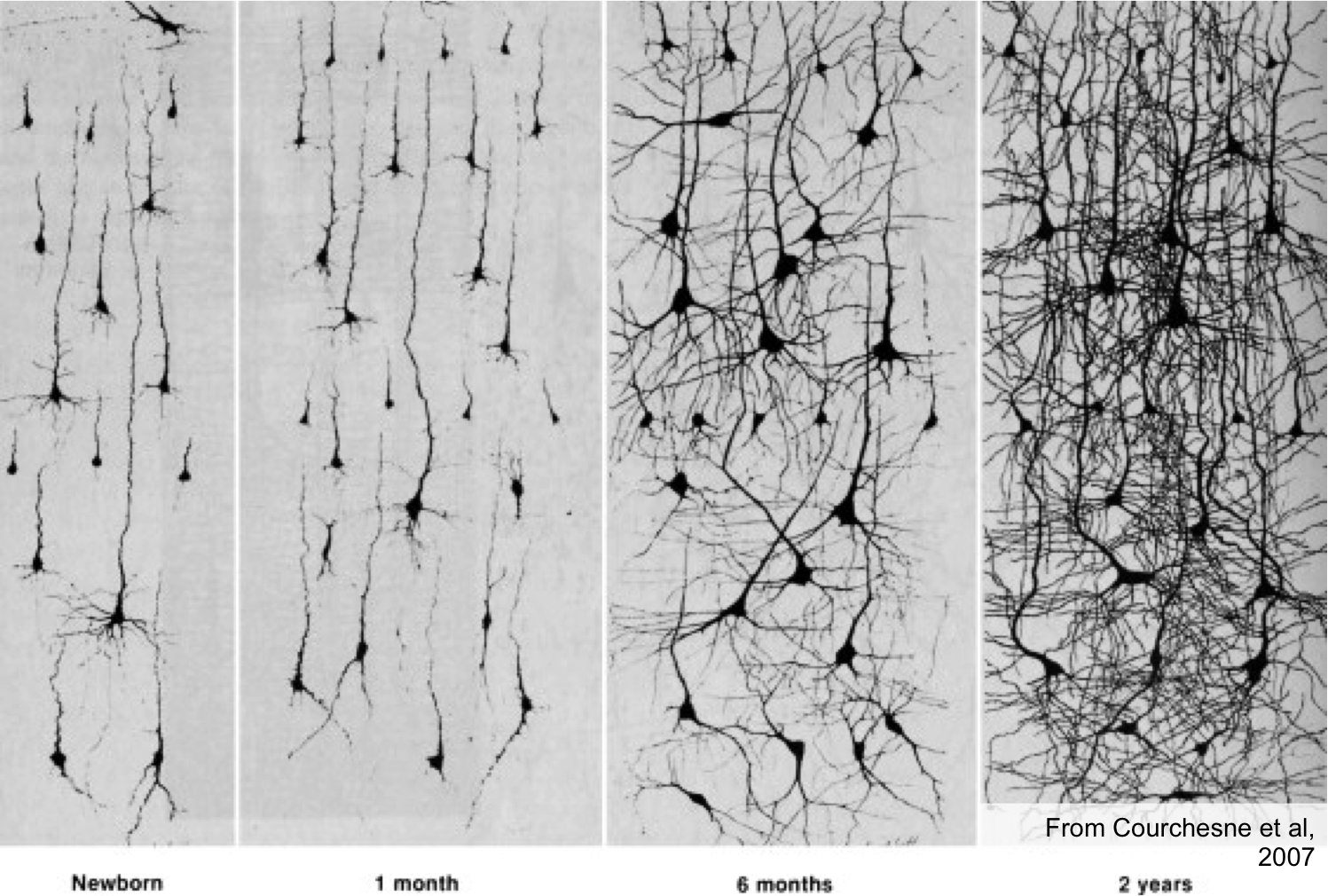
golgistainbirthto2years Sapien Labs Neuroscience Human Brain Diversity Project
Golgi needed a staining technique to reveal neurons in their entire, undamaged form to confirm his reticular theory. Reticular theory stated that the entire nervous system was a continuous network of cells without gaps or synapses in between the cells.
6 Views of a Neuron by Golgi and Cajal Breakthroughs
Golgi staining is a classical technique based on a deposition of metal precipitate in a random set of neurons. Despite their versatility, Golgi methods have limitations that largely precluded.

Representative higher magnification images of Golgi staining done in... Download Scientific
The Golgi-Cox staining method is a cost-effective, relatively simple means of staining a random sample of neurons within the brain. First developed by Golgi 1 and modified by Cox 2 in the 1800s, researchers have further refined this technique over the years to produce clear, well-stained neurons that can be used to visualize and quantify both.

Photomicrographs showing of different types of Golgi staining of... Download Scientific Diagram
Discovered already by Golgi ( 1873 ), the non-invasive Golgi staining method is far from out-of-date, and it facilitates an analysis of neuronal morphology with axonal and dendritic arborization and spines through visualization of only a low percentage of neurons (1-3%).
