Sections of the Lungs Seattle Cancer Care Alliance
Lung Diagram Labeled EdrawMax Template
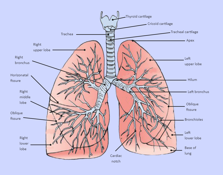
Labeled Diagram of the Human Lungs Lungs are an excellent example of how several tissues can be compactly arranged, yet providing a large surface area for gaseous exchange. The current article provides a labeled diagram of the human lungs as well as a description of the parts and their functions.

Foods to help keep your lungs and respiratory system healthy
Overview A step-by-step explanation of how your lungs work. What are your lungs? Your lungs make up a large part of your respiratory system, which is the network of organs and tissues that allow you to breathe. You have two lungs, one on each side of your chest, which is also called the thorax.
CrossFit Anatomy of the Lungs
A medical diagram showing the lobes of the lungs (organ of the respiratory system) with text labels. Vector graphic. diagram of the respiratory system with labels stock illustrations lung. The lungs are the primary organs of respiration in humans and many other animals including a few fish and some snails.
The lungs Lung cancer Macmillan Cancer Support
Introduction This e-Anatomy module is dedicated to the anatomy of the thorax (lungs, pleura, heart, aorta, thoracic lymph nodes and other relevant anatomical structures) using a normal thorax CT.

A schematic drawing of the lungs and airway tree in which several
ISSN 2534-5079. We created an anatomy atlas of the chest and the mediastinum which is an interactive tool for studying the cross-sectional anatomy of the normal thorax based on an enhanced multidetector computed tomography with helical angiography of the thorax (axial plane). Anatomical structures are visible as interactive labeled images.

Lung Structure BioNinja
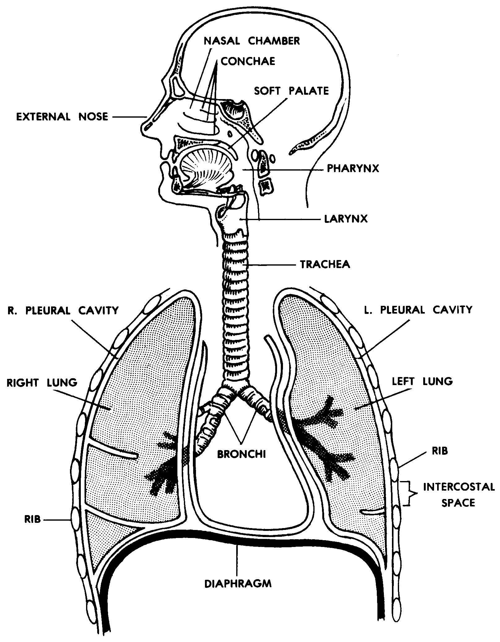
The respiratory system, also called the pulmonary system, consists of several organs that function as a whole to oxygenate the body through the process of respiration (breathing). This process involves inhaling air and conducting it to the lungs where gas exchange occurs, in which oxygen is extracted from the air, and carbon dioxide expelled.
Images 07. Respiratory System and Breathing Basic Human Anatomy
Browse 170+ diagram of the respiratory system with labels stock photos and images available, or start a new search to explore more stock photos and images. Sort by: Most popular The respiratory system The human respiratory system medical illustration with internal organs lung.
Lung Anatomy & Function Lung Nodule, Lung Disease and Lung Infection
1 /14. Your lungs work all day and night, whether you're awake or asleep. That's 20,000 or so breaths per day! By the time you're 50, you have taken around 400 million breaths. Diseases like.

Pin on A&P.4.Heart.Lung
Anatomy Function Associated Conditions Tests The lungs are a major organ that is part of the respiratory system, taking in fresh air and getting rid of old, stale air. This mechanism of breathing also helps to allow you to talk. By taking in fresh air, the lungs are able to help oxygenate blood to be carried around your body.

Medical Education Chart of Biology for Lungs Diagram. Vector
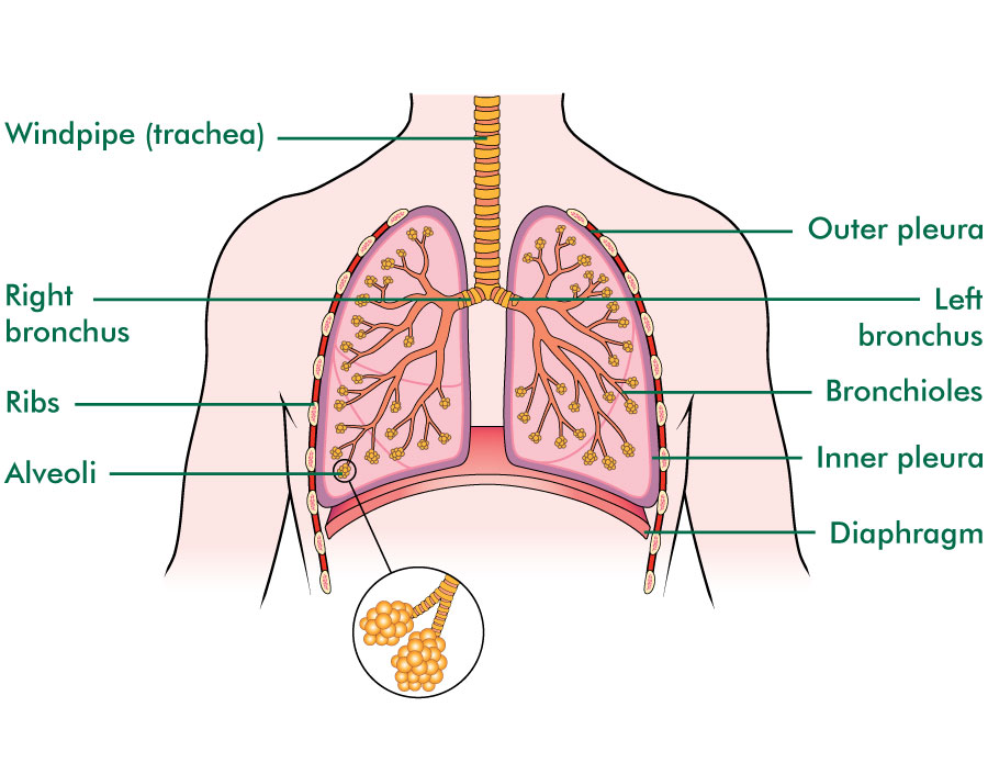
The lungs' main role is to deliver oxygen to the blood and remove carbon dioxide from the blood. Air enters the nose or mouth and passes through your windpipe and into the bronchial tubes when you breathe in. The bronchial tubes lead into the lungs and branch out into smaller tubes known as bronchioles, which end in small air sacs known as alveoli.

The lungs, trachea and bronchi, mediastinum and detail of chest wall
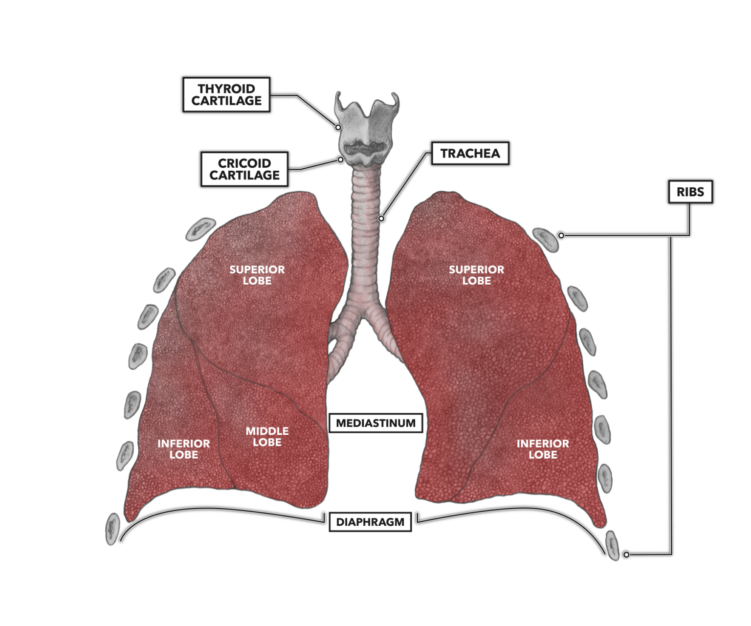
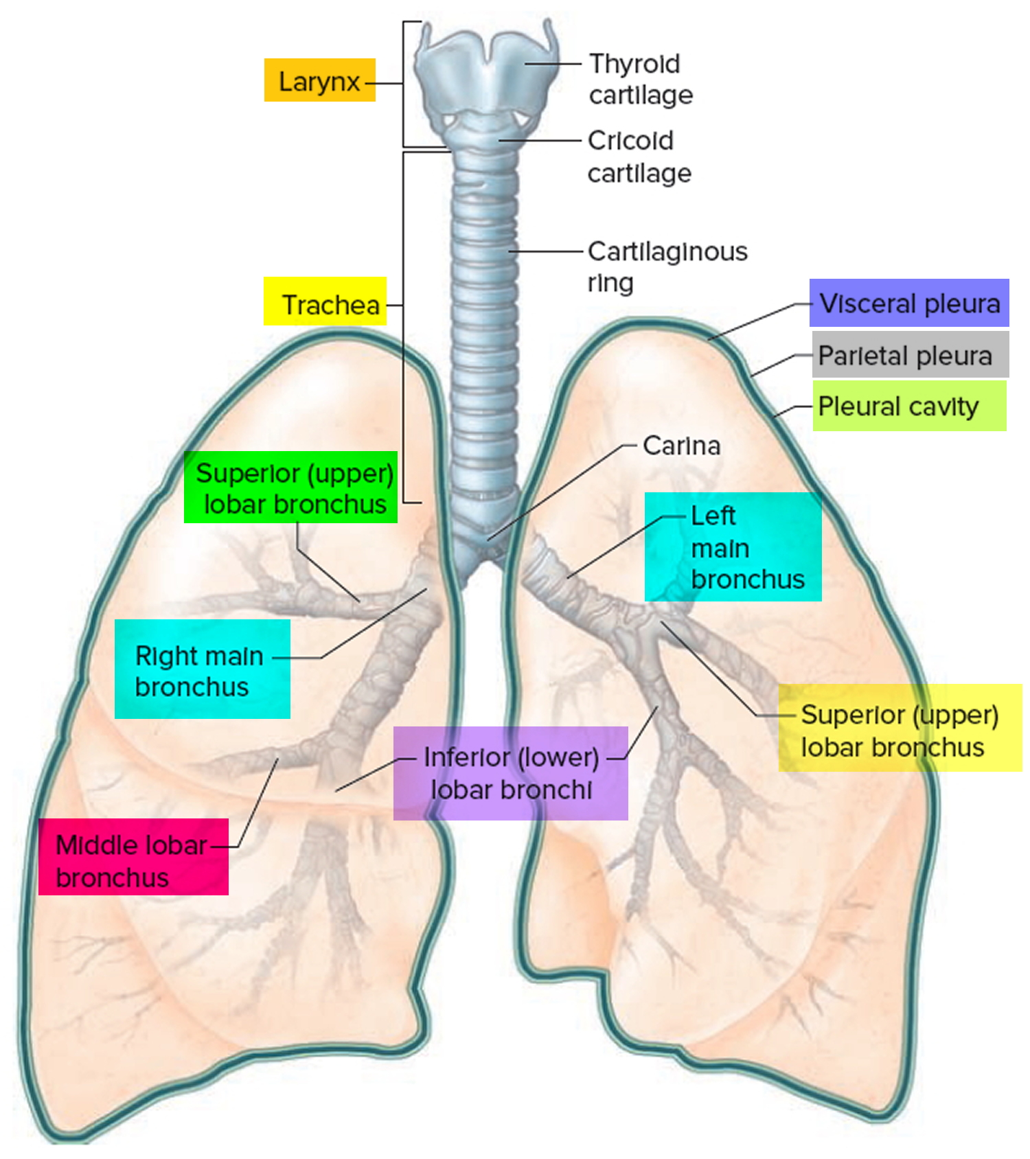
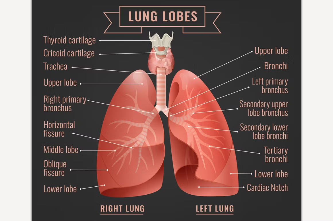
Functionally, the lung is divided into a series of bronchopulmonary segments. The bronchopulmonary segments are the largest subdivision of a lobe. They are separated from adjacent segments by connective tissue septa and are also surgically resectable. They are 10 bronchopulmonary segments in the left lung and 8-10 in the left lung [9].
Pleural effusion causes, types, symptoms, diagnosis and treatment
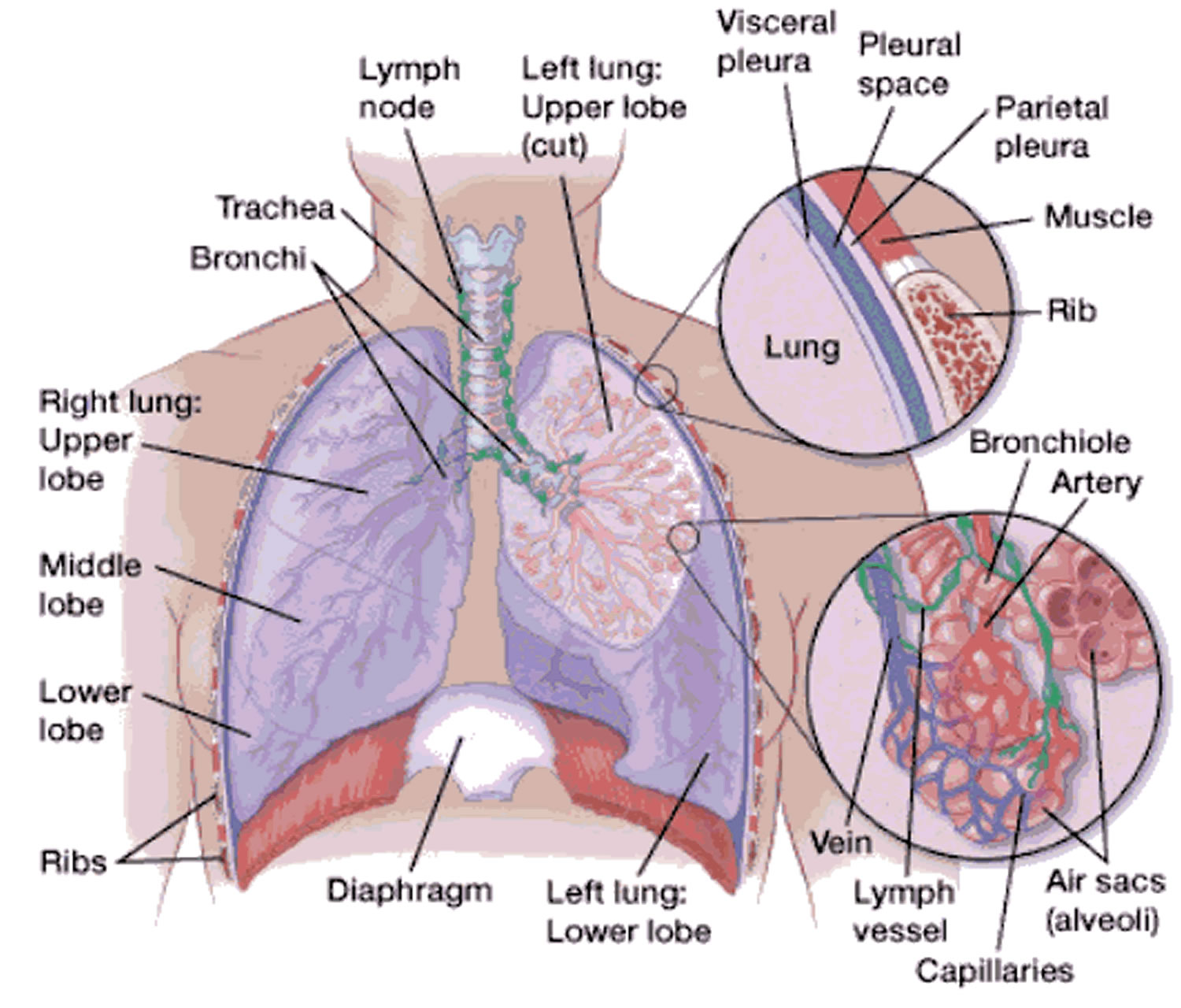
Blood supply. The lungs have dual, parallel blood supply referred to as pulmonary and systemic circuits. The pulmonary circuit arises from the heart and brings deoxygenated blood to the lung for gas exchange. This circuit also takes oxygenated blood back to the heart to be redistributed to the rest of the body.

Respiratory system the lung association Respiratory system, Human
Blood Supply The major function of the lungs is to perform gas exchange, which requires blood from the pulmonary circulation. This blood supply contains deoxygenated blood and travels to the lungs where erythrocytes, also known as red blood cells, pick up oxygen to be transported to tissues throughout the body.
Human lungs infographic Education Illustrations Creative Market
The superior end of the lungs narrows to a rounded tip known as the apex. The inferior end of the lungs, known as the base, rests on the dome-shaped diaphragm. The base of the lungs is concave to follow the contour of the diaphragm. The left lung is slightly smaller than the right lung because 2/3 of the heart is located on the left side of the.
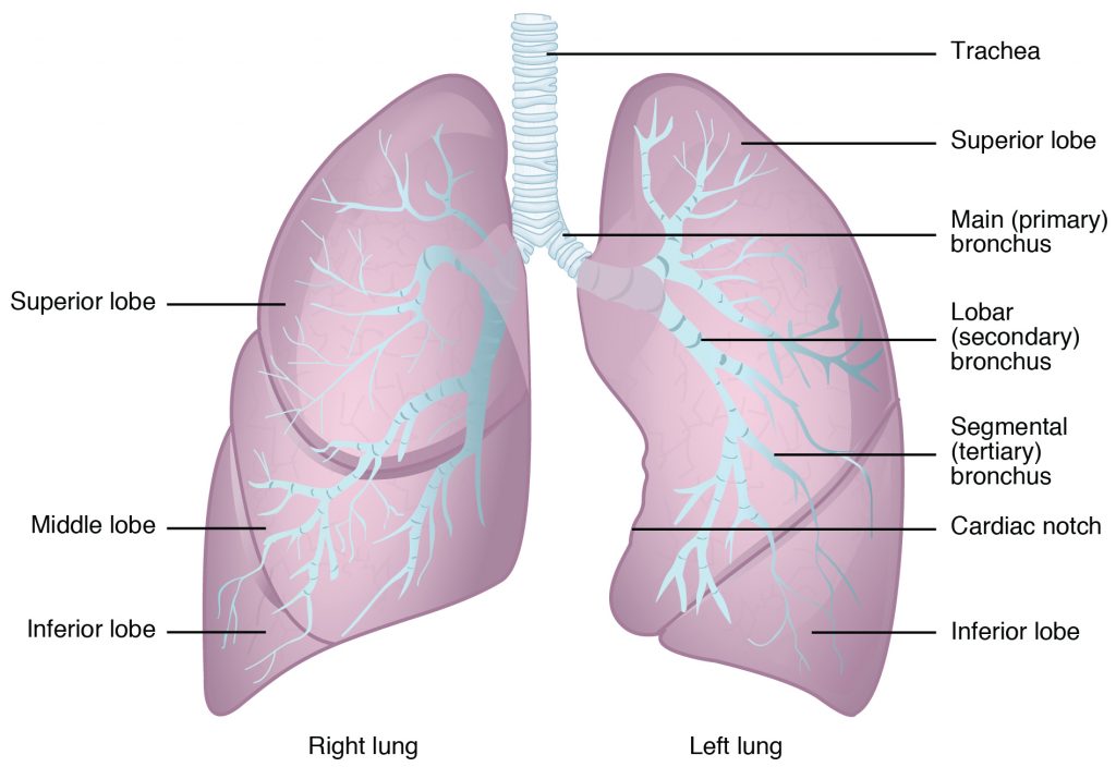
Diagram Of The Lungs With Labels Labeling Of The Lungs Label The Lungs
Respiratory system This chart of the RESPIRATORY SYSTEM shows how you breathe. Breathing is the process that brings oxygen in the air into your lungs and moves oxygen and through your body. Our lungs remove the oxygen and pass it through our bloodstream, where it's carried off to the tissues and organs that allow us to walk, talk, and move.

Human Lungs Infographic Stock Illustration Download Image Now iStock
Respiration or the act of breathing, is an automatic process, regulated by the brain. In this article, we shall discuss lungs in detail. Simple Lungs diagram The lung, the human gas-exchanging structure, resides in the chest (thorax) wherein its intricate tissues are protected by the muscular and bony thoracic cage. Structure of the Lungs
