[DIAGRAM] Diagram Of Cytosol

animal cell diagram easy Kris Hammett
Diagram of a typical plant cell: Diagram of a plant cell with components labeled. Image modified from OpenStax Biology. Both animal and plant cells have mitochondria, but only plant cells have chloroplasts. Plants don't get their sugar from eating food, so they need to make sugar from sunlight. This process (photosynthesis) takes place in the.

Plant Cell Diagram Wallpapers Power
1. Prokaryotes are the simplest cells without a nucleus and cell organelles. 2. Prokaryotic cells are the smallest cells (1-10 μm). 3. Unicellular and earliest to evolve (~4 billion years ago), still available. 4. The cell wall is rigid. ADVERTISEMENTS: 5. These cells reproduce asexually. 6. They include bacteria and archaea. 7.

Explain the nucleus of a cell with a neat labeled diagram Science
A Labeled Diagram of the Animal Cell and its Organelles There are two types of cells - Prokaryotic and Eucaryotic. Eukaryotic cells are larger, more complex, and have evolved more recently than prokaryotes. Where, prokaryotes are just bacteria and archaea, eukaryotes are literally everything else.
Human Cell Diagram 6406474 Vector Art at Vecteezy
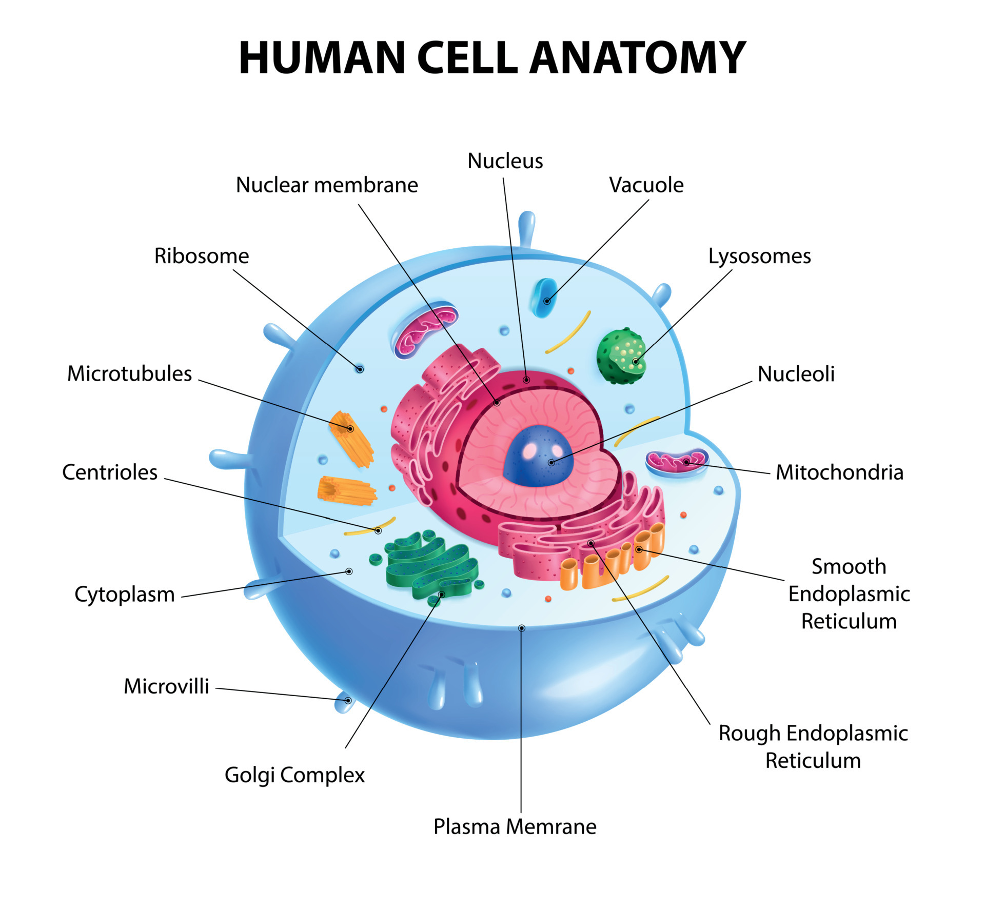
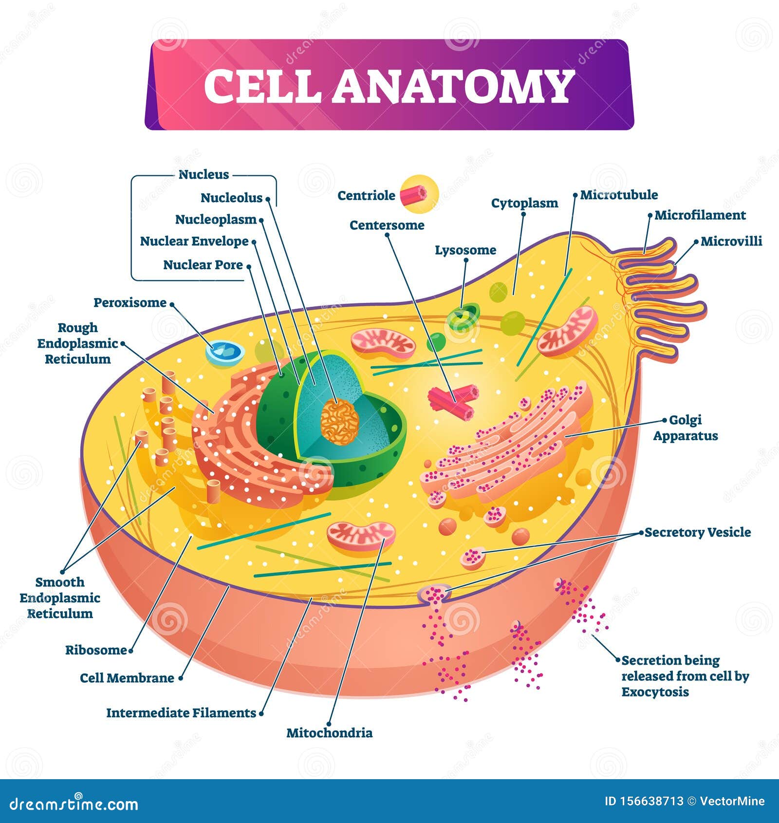
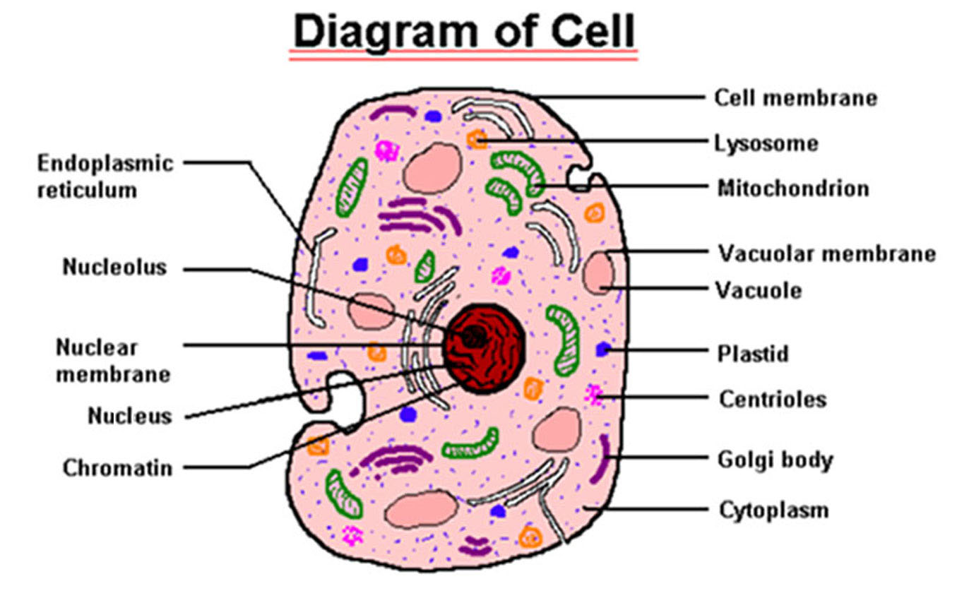
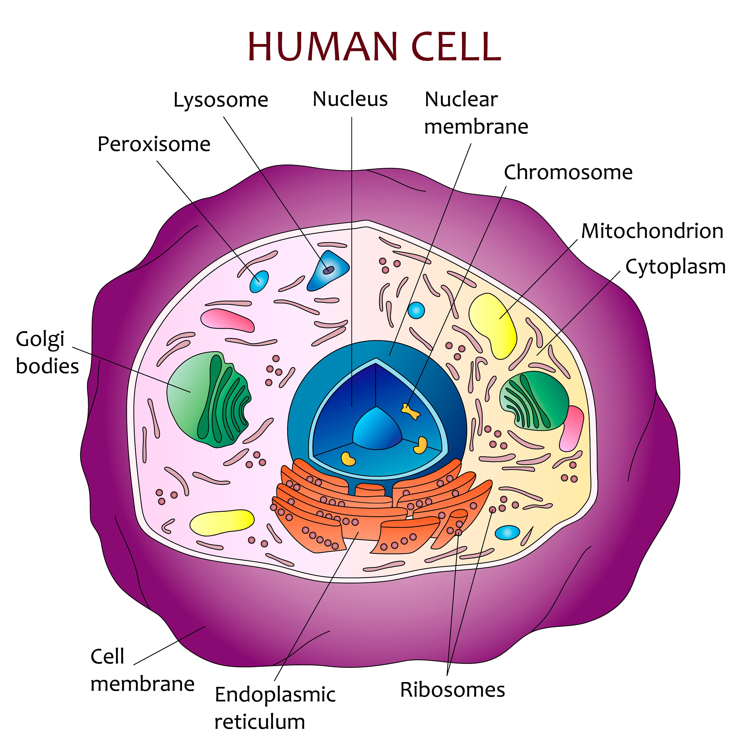
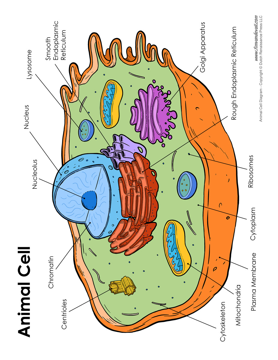
The well labelled diagram of an animal cell consists of all the organelles and the structural components of an animal cell. An animal cell is the basic unit of any living animal. All the cells found in any living animal are made up of similar components and organelles and are eukaryotic cells. Hence, a well labelled diagram of animal cells will be quite uniform amongst all the different.

Cell Diagram Labeled Simple SexiezPicz Web Porn
cell, in biology, the basic membrane-bound unit that contains the fundamental molecules of life and of which all living things are composed.A single cell is often a complete organism in itself, such as a bacterium or yeast.Other cells acquire specialized functions as they mature. These cells cooperate with other specialized cells and become the building blocks of large multicellular organisms.
Cell Anatomy (3D Labeled Diagram) Science
I've created two interactive diagrams for an upcoming open textbook for high-school level biology. The cell structure illustrations for these diagrams were generated in BioRender. Both diagrams feature a drag-and-drop labelling activity created with H5P here on Learnful. These h5p resources are made available openly with the CC BY license.

Labeled Animal Cell Diagram
Diagram Of Animal Cell Animal cells are eukaryotic cells that contain a membrane-bound nucleus. They are different from plant cells in that they do contain cell walls and chloroplast. The animal cell diagram is widely asked in Class 10 and 12 examinations and is beneficial to understand the structure and functions of an animal.
Cell Anatomy Vector Illustration. Labeled Educational Structure Diagram
Animal Cell Anatomy. The cell is the basic unit of life. All organisms are made up of cells (or in some cases, a single cell). Most cells are very small; in fact, most are invisible without using a microscope. Cells are covered by a cell membrane and come in many different shapes.
Education 645 High School Biology
Animal cells are eukaryotic cells, meaning they possess a nucleus and other membrane-bound organelles. Unlike plant cells, animal cells do not have cell walls, allowing for more flexibility in shape and movement. A plasma membrane encloses the cell contents of both plant and animal cells, but it is the outer coating of an animal cell.
Human cell diagram Etsy
Animal Cell: Structure, Parts, Functions, Labeled Diagram June 6, 2023 by Faith Mokobi Edited By: Sagar Aryal An animal cell is a eukaryotic cell that lacks a cell wall, and it is enclosed by the plasma membrane. The cell organelles are enclosed by the plasma membrane including the cell nucleus.

Animal Cell Diagram Organelles Simple Functions And Diagram Gambaran
Key points: All cells have a cell membrane that separates the inside and the outside of the cell, and controls what goes in and comes out. The cell membrane surrounds a cell's cytoplasm, which is a jelly-like substance containing the cell's parts. Cells contain parts called organelles. Each organelle carries out a specific function in the cell.

FileCell Structure , Cell Diagram.png Wikimedia Commons
Biology library 37 units · 127 skills Unit 1 Intro to biology Unit 2 Chemistry of life Unit 3 Water, acids, and bases Unit 4 Properties of carbon Unit 5 Macromolecules Unit 6 Elements of life Unit 7 Energy and enzymes Unit 8 Structure of a cell Unit 9 More about cells Unit 10 Membranes and transport Unit 11 More about membranes

South Pontotoc Biology Plant and Animal Cell Diagrams
The plasma (cell) membrane separates the inner environment of a cell from the extracellular fluid. It is composed of a fluid phospholipid bilayer (two layers of phospholipids) as shown in figure 4.1.2 4.1. 2 below, and other molecules. Not many substances can cross the phospholipid bilayer, so it serves to separate the inside of the cell from.

What is a cell? Facts
Labeled diagram of a typical animal cell Nucleus. The nucleus contains all the genetic material in a cell. This genetic information is called deoxyribonucleic acid (DNA). DNA contains all the instructions for making proteins, which control all of the body's activities. Therefore, the nucleus is like the manager's office of the cell.

Cell Structure
Cell diagram labeled Cell diagram unlabeled Learn faster with interactive cell quizzes Sources + Show all What are the parts of a cell? There exist two general classes of cells: Prokaryotic cells: Simple, self-sustaining cells (bacteria and archaea) Eukaryotic cells: Complex, non self-sustaining cells (found in animals, plants, algae and fungi)
Animal Cell Model Diagram Labeled / Animal Cell Model Diagram Project
What exactly is its job? The plasma membrane not only defines the borders of the cell, but also allows the cell to interact with its environment in a controlled way. Cells must be able to exclude, take in, and excrete various substances, all in specific amounts.
