Shoulder Xray Interpretation Radiology Geeky Medics
Citation, DOI, disclosures and article data. Normal radiographic measurements of the shoulder are important in the evaluation of the osseous relationships in plain radiography. Normal measurements do not rule out pathology and must be considered in the context of other findings and the clinical presentation. acromioclavicular (AC) joint space.

X Ray Normal Shoulder
Shoulder radiographs are often the only imaging exam necessary for the evaluation of acute shoulder trauma, calcific tendonitis, arthritis, and osteolysis of distal clavicle (in athletes) [ 1 ]. Computed tomography — Computed tomography (CT) of the shoulder is usually reserved for evaluation of fracture/fracture-dislocation or for a.
Normal Shoulder, Xray Photograph by Du Cane Medical Imaging Ltd
Murphy A, Normal shoulder radiographs. Case study, Radiopaedia.org (Accessed on 09 Jan 2024) https://doi.org/10.53347/rID-53052

real shoulder xray anatomy Diagram Quizlet
The vast majority of people believe that shoulder X-rays are a good way to determine what's causing shoulder pain. Stress, among other things, can make your shoulder muscles quite cranky. Many doctors also say that an X-ray is a good way to investigate shoulder pain. They might find things like medial acromial and lateral clavicular sclerosis.
Shoulder Xrays The Bone School
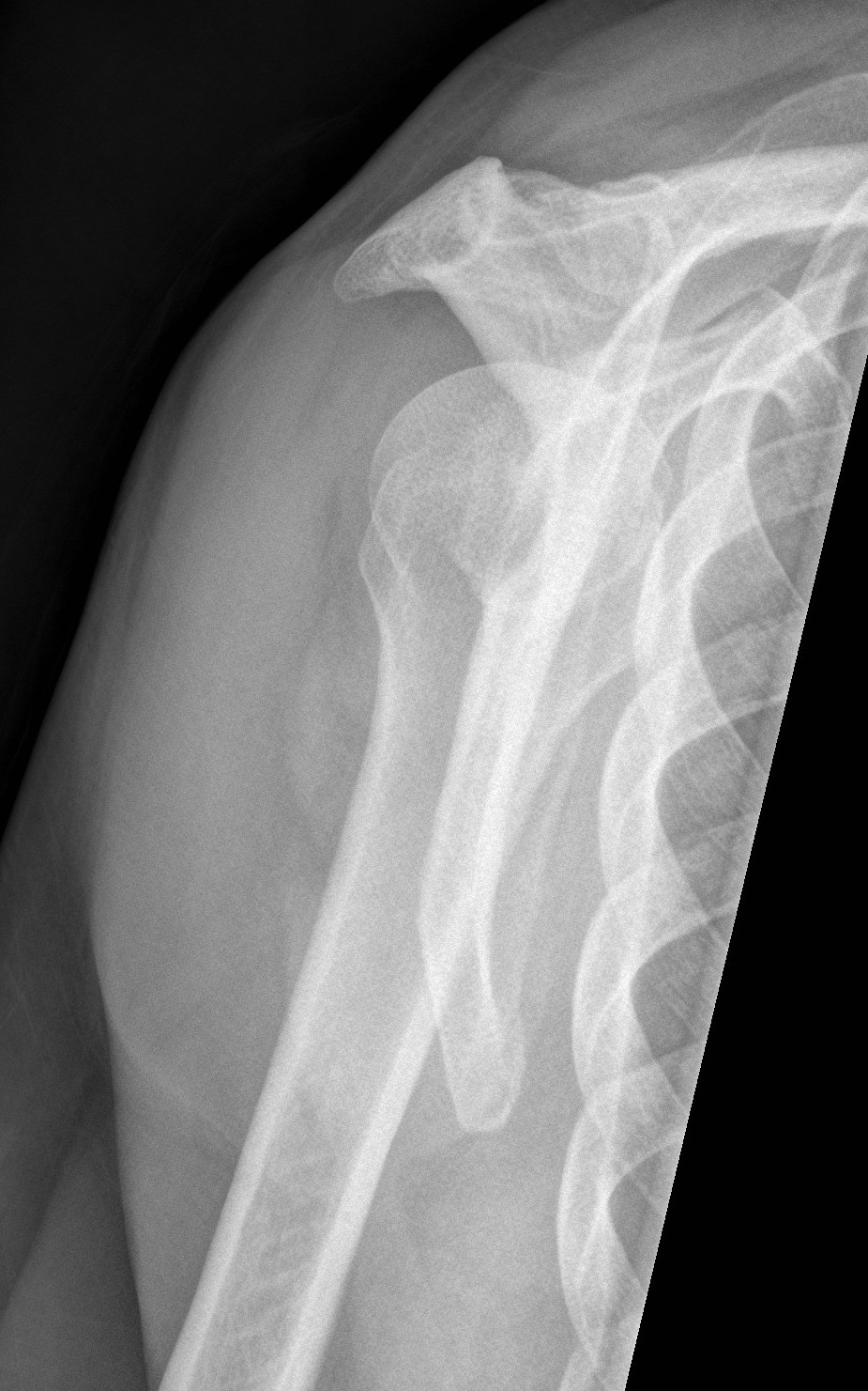
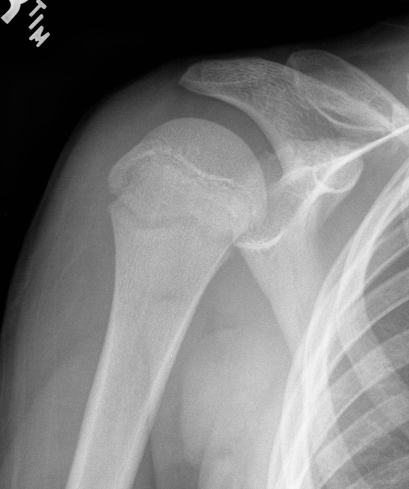
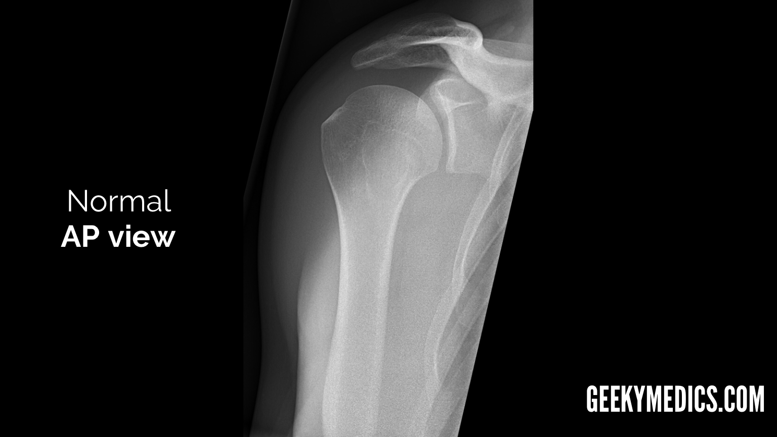
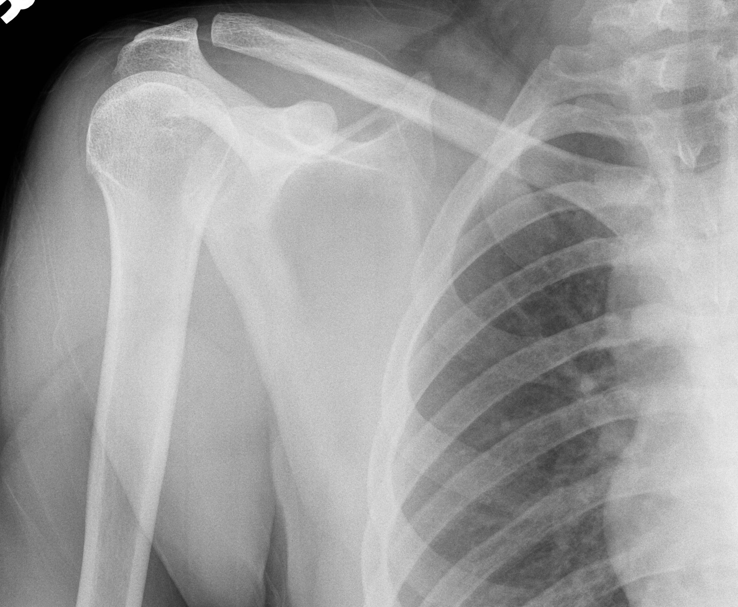
Fig. 3.1. Anteroposterior shoulder radiograph. While achieving anteroposterior shoulder X-ray in neutral position, the patient is erect or in supine position. Central X-ray should be directed to 2.5 cm inferior to the coracoid process. Anteroposterior shoulder view allows assessment of especially the humeral head lesions and clavicular fractures.
NORMAL SHOULDER 1
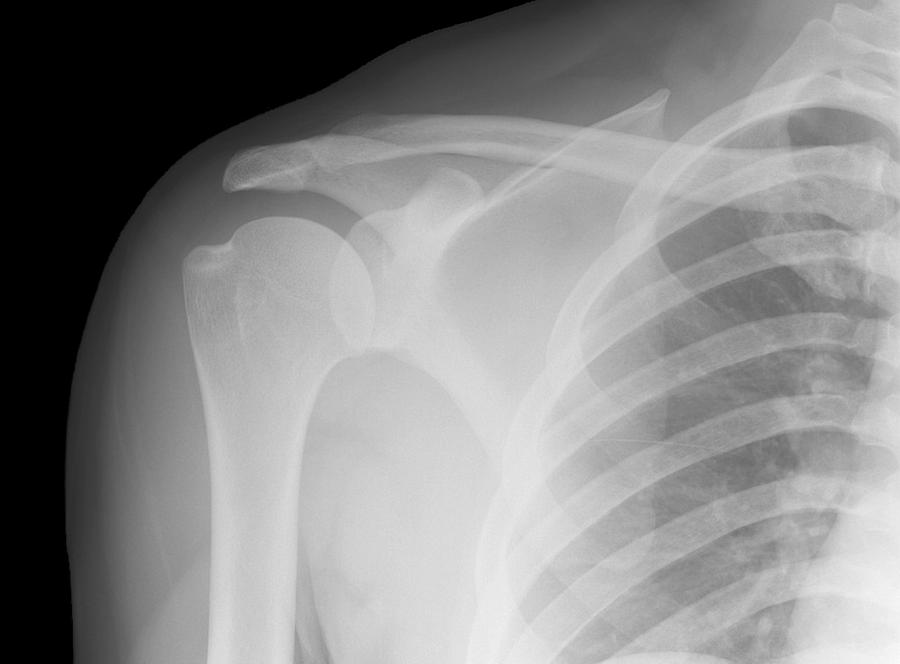
This projection is a true anterior-posterior (AP) view of the shoulder. The Grashey view involves angling the beam laterally or rotating the patient posteriorly(2). These adjustments remove the view of the overlap between the humerus and the glenoid. The removal allows better evaluation of joint congruity, humeral head subluxation, and the.

Normal shoulder Image
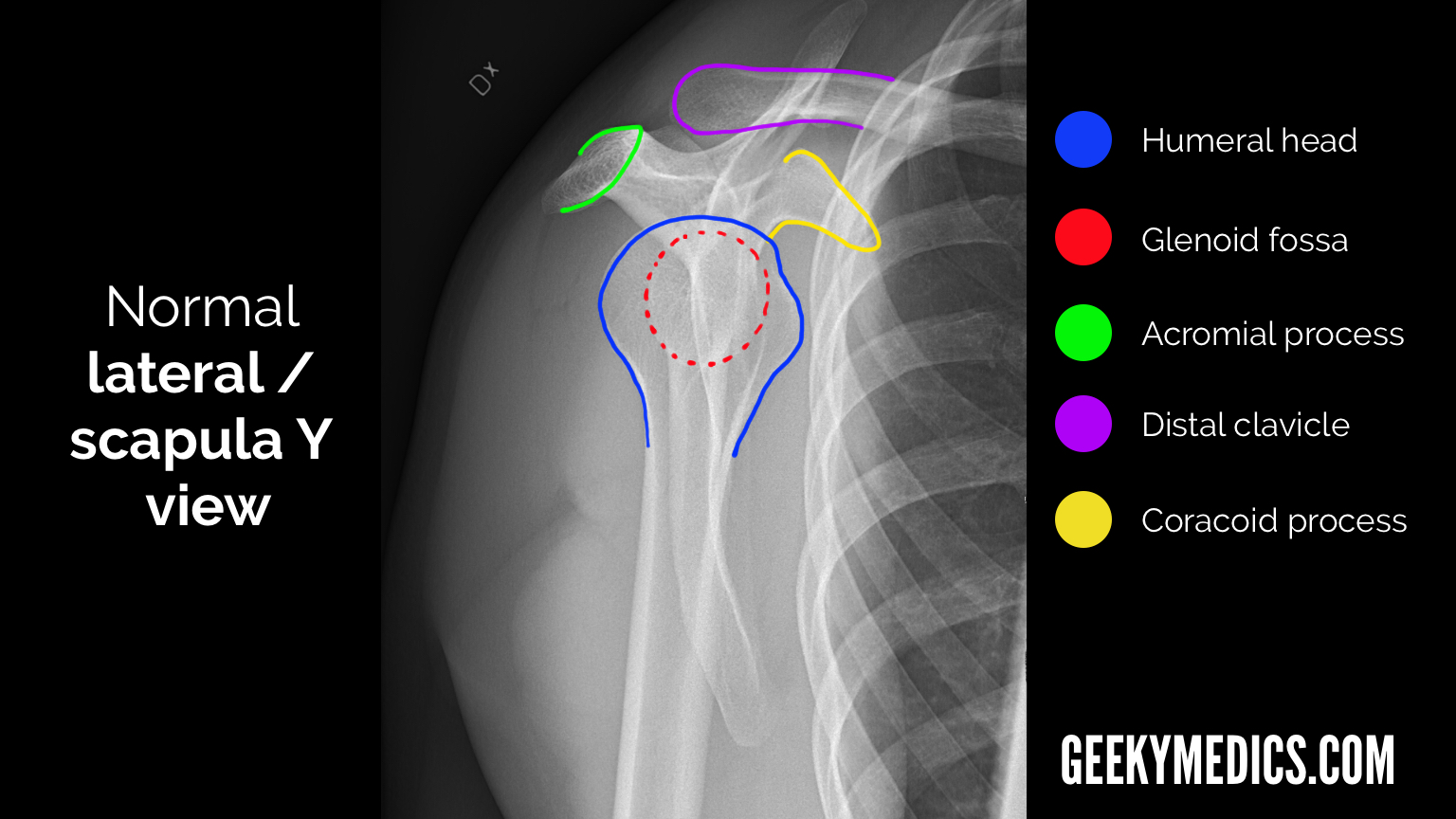
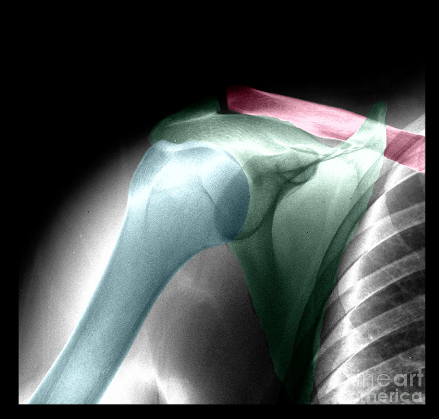
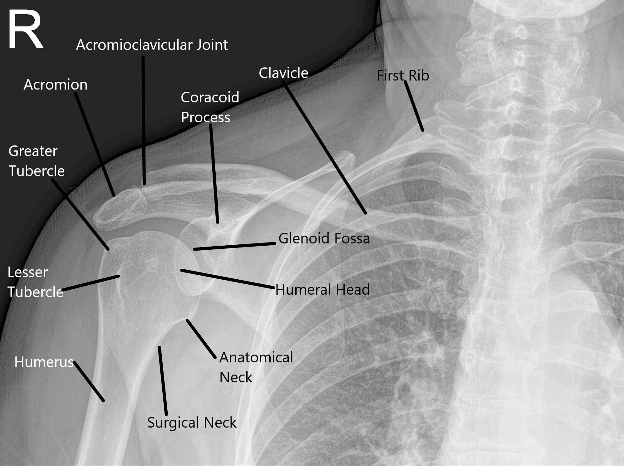
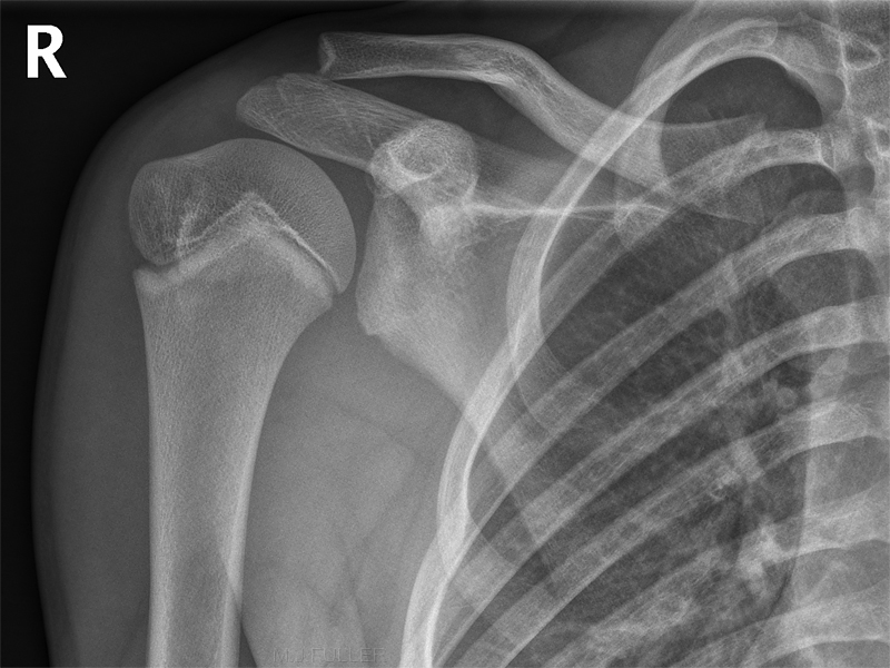
Citation, DOI, disclosures and case data. Scroll or drag your finger down to reveal the radiographic anatomy for each shoulder view. The normal osseous anatomy is outlined.
Normal Shoulder, Xray Photograph by Living Art Enterprises
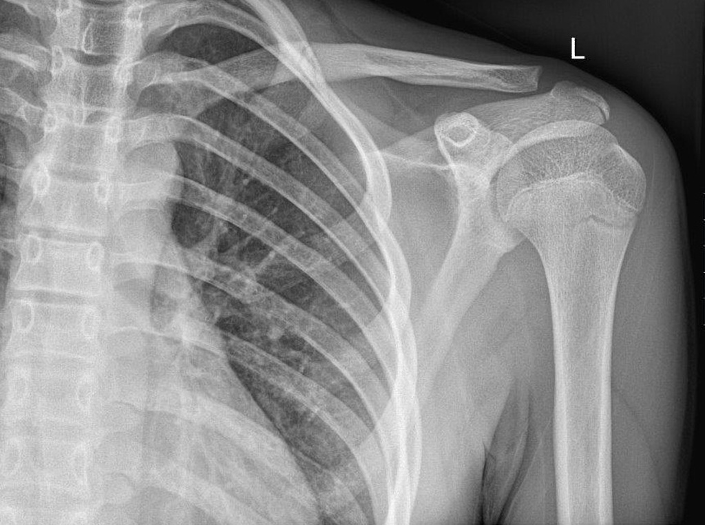
Citation, DOI, disclosures and article data. The shoulder series is fundamentally composed of two orthogonal views of the glenohumeral joint including the entire scapula. The extension of the shoulder series depends on the radiography department protocols and the clinical indications for imaging.
UCSD Musculoskeletal Radiology
The shoulder is passively abducted in the scapular plane to 90°. The examiner's other hand is placed over the patient's shoulder overlying the anterior acromion and greater tuberosity. The examiner passively internally and externally rotates the shoulder detecting the presence of palpable crepitus.

437.1 shoulder NorMal1 Normal shoulder Xray series Flickr
glenoid version for total shoulder arthroplasty. Magnetic Resonance Imaging. Overview. MRI is best for evaluating soft tissue structures and evaluating bone contusions or trabelcular microfractures. the stronger the magnet, the higher the intrinsic signal-to-noise ratio (e.g. a 3 Tesla MRI machine has 9x the proton energy of a 1.5 Tesla MRI.

Normal shoulder, Xray Stock Image F003/9192 Science Photo Library
Introduction. An X-ray of the shoulder is a frequently conducted examination and is mainly used for diagnosing a fracture. Some of the key topics are proximal humeral fracture, shoulder dislocation, Bankart lesion and osteoarthritis. KEY TOPICS/TERMS: Proximal humeral fracture. Shoulder dislocation. Hill-Sachs lesion.
Shoulder Xray Interpretation Radiology Geeky Medics
Case Discussion. Additional to joint alignment and fractures, shoulder radiographs should be assessed for rotator cuff calcification, as it can present with acute shoulder pain.
NORMAL SHOULDER 5
Posterior shoulder dislocation. less than 5% of glenohumeral dislocations but often overlooked. common in adults following a seizure or in the elderly. humeral head forced posteriorly in internal rotation whilst arm is abducted. classically, the humeral head is rounded on AP - light bulb sign. associated with anteromedial fracture of humeral head.
UCSD Musculoskeletal Radiology
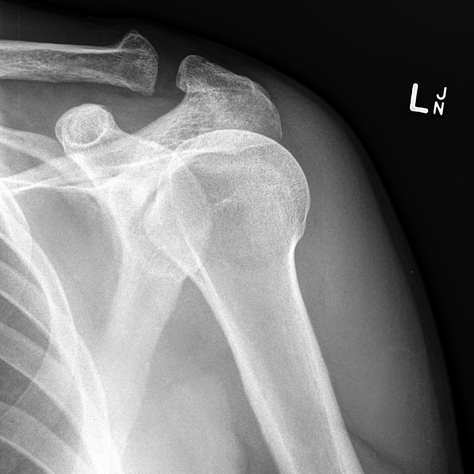
Your shoulder joint can move in more directions than any other joint in your body. A normal shoulder X-ray will show the bones that make up this ball-and-socket joint: Humerus (upper arm bone). Scapula (shoulder blade), which connects to the humerus. Acromion (a piece of bone that projects off the scapula). Clavicle (collarbone), which connects.
Arthroscopy Shoulder Joint Complete Orthopedics Multiple NY Locations
Look for disruption or a buckle in the cortex or any fracture fragments. They should all be smooth. The clavicle is a good bone to start with - it is by far the most common paediatric shoulder injury. Midshaft fractures account for 80% of clavicle fractures. Make sure there are no distal or medial fractures as they can often be subtle.
NORMAL CHILD SHOULDER
Gaillard F, Normal shoulder. Case study, Radiopaedia.org (Accessed on 12 Jan 2024) https://doi.org/10.53347/rID-7505
