Palmar view of hand anatomy including tendons and pulleys Hand anatomy, Anatomy images, Anatomy

Trigger Finger Fort Worth, TX The Hand to Shoulder Center
Many studies have looked at which combination of pulleys are required for mechanically efficient finger flexion and their results have guided hand surgeons in pulley preservation and reconstruction. In this article we review the anatomy of the flexor pulley system and its clinical relevance.

Pulley Injuries Explained (Part 1) — Grassroots Physical Therapy
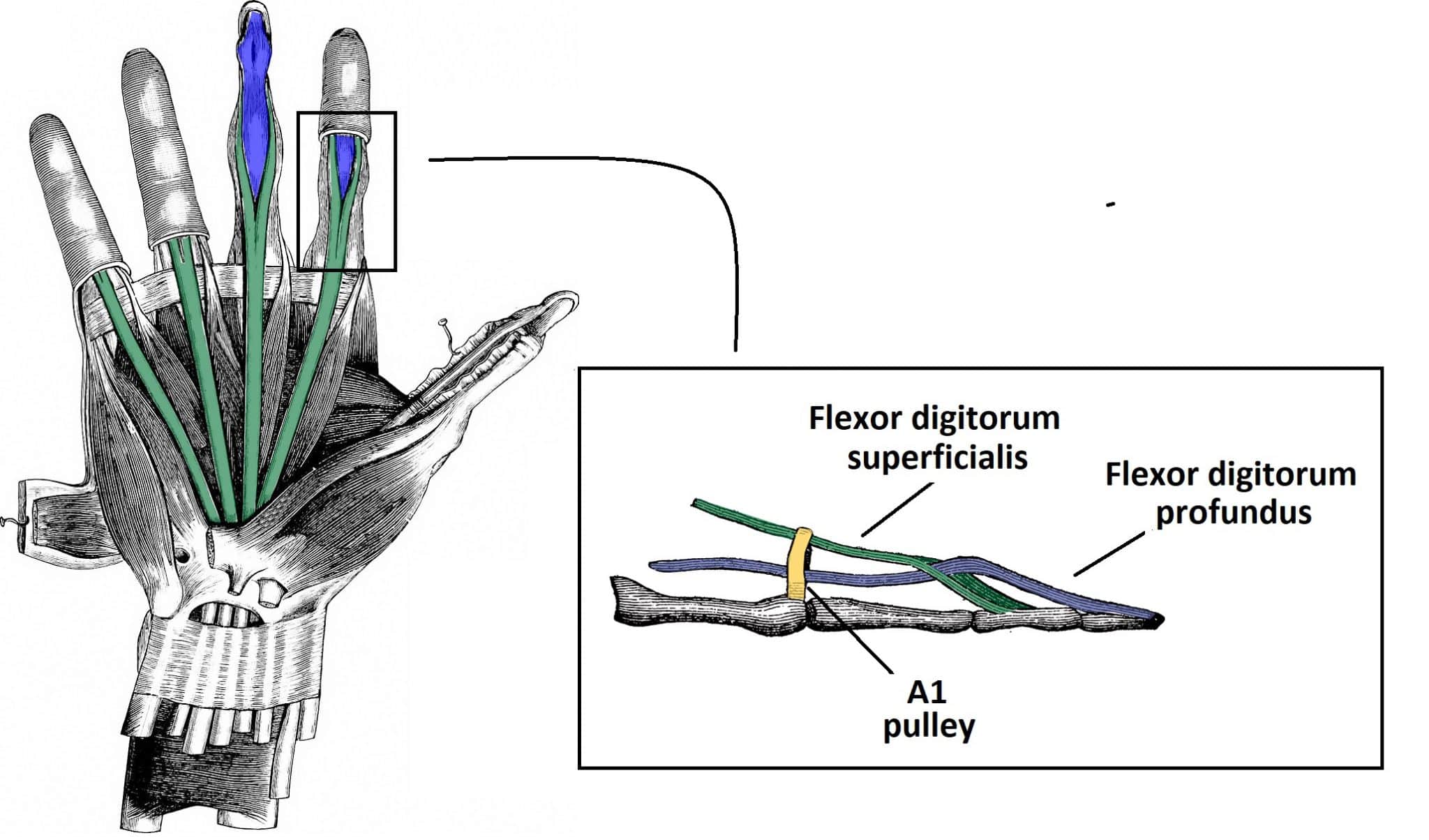
The pulleys are fibrous tissue condensations, which almost encircle the flexor tendons forming a fibro-osseous channel that functions to keep the tendons adjacent to the phalanges. This enables the transfer of a translational force generated from the muscle-tendon unit into a rotational moment on the phalanges.

Wrist and hand Basicmedical Key
Detail the anatomy and biomechanics of the flexor pulley system. Describe the epidemiology and etiology of flexor pulley system injuries. Recognize the clinical manifestations of closed pulley system rupture. Offer diagnostic strategies for various types of closed pulley system ruptures.

Flexor Pulley System Anatomy, Injury, and Management Journal of Hand Surgery
Pulley Lesion of the Fingers Mark Awh, M.D. Clinical History: A 28 year-old rock climber presents with persistent pain and swelling at the index finger. (1a) A T2-weighted sagittal image with fat-suppression and (1b) a proton density-weighted fat-suppressed axial image are provided. What are the findings" What is your diagnosis? 1a 1b Figure 1:
The Flexor Pulley System of the Hand Annular Cruciate Oblique TeachMeAnatomy
Doyle JR. Anatomy and function of the palmar aponeurosis pulley. J Hand Surg [Am]. 1990 Jan. 15(1):78-82. [QxMD MEDLINE Link]. Doyle JR, Blythe WF. Anatomy of the flexor tendon sheath and pulleys of the thumb. J Hand Surg [Am]. 1977 Mar. 2(2):149-51. [QxMD MEDLINE Link]. Bunnell S. Repair of tendons in the fingers and description of two new.

Palmar view of hand anatomy including tendons and pulleys Hand anatomy, Anatomy images, Anatomy
summary. Flexor Tendon Injuries are traumatic injuries to the flexor digitorum superficialis and flexor digitorum profundus tendons that can be caused by laceration or trauma. Diagnosis is made clinically by observing the resting posture of the hand to assess the digital cascade and the absence of the tenodesis effect.
The Flexor Pulley System of the Hand Annular Cruciate Oblique TeachMeAnatomy
A thorough awareness of hand and upper limb anatomy is vital to understanding injuries to these regions, planning subsequent management, and anticipating complications. We will review anatomy here systematically.. Pulleys are either cruciate and annular (Fig. 1.8). Annular pulleys function to prevent bowstringing.

Alila Medical Media Finger tendon sheath and pulleys labeled. Medical illustration
Functional Anatomy of the Hand Introduction The hand contains a number of joints that allow complex actions, such as manipulating, gripping and grasping objects. Optimal hand function requires adequate strength, sensation, range of motion, and dexterity.

Varied Anatomy of the Thumb Pulley System Implications for Successful Trigger Thumb Release
A trigger finger, sometimes referred to as a trigger thumb or stenosing tenosynovitis, can occur if one of three things happen: 1. The tendon enlarges (does not fit through pulley well); 2. The lining increases in thickness (does not fit through pulley well); 3. the pulley becomes thicker (the opening for the tendon gets smaller).

Physio Health Finger and Hand Anatomy, and Grip
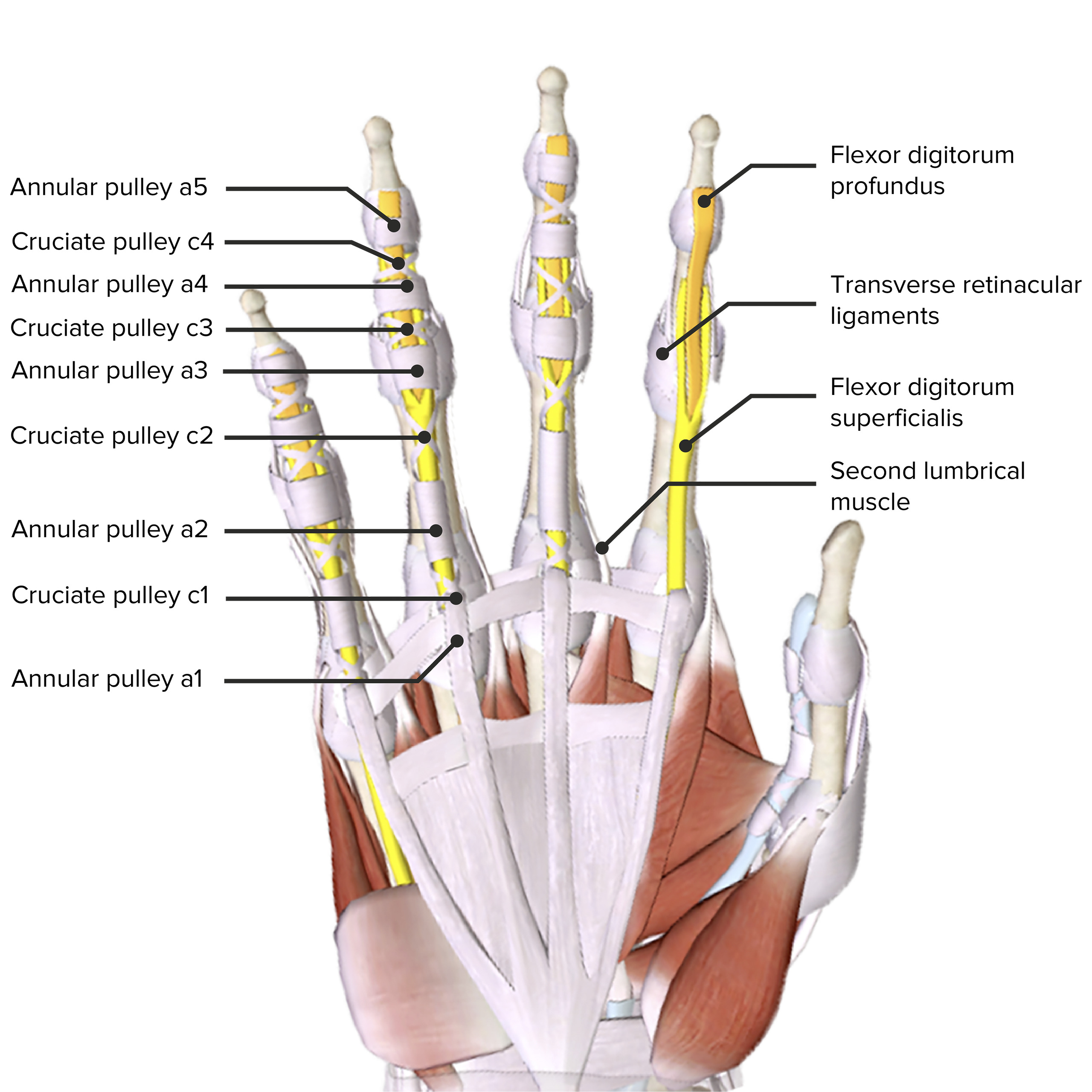
The cruciate pulleys, allows the flexor sheath to concertina, that is to change length during flexion-extension. The thumb has an AI pulley volar to the MP joint and an A2 pulley volar to the IP joint. The oblique pulley is the largest and most important pulley in the thumb and lies between A1 and A2 obliquely across the proximal phalanx.
Hand Anatomy Concise Medical Knowledge
From Lin GT, Amadio PC, Cooney WP: Anatomy of human digital flexor pulleys (accepted for publication by J Hand Surg). flGURE 3. The cruciate pulleys have one fixed attachment, to bone and one moveable attachment, to volar plate. From Lin GT, Amadio PC, Cooney WP: Anatomy of human digital flexor pulleys (accepted for publication by J Ha~d Surg).
.jpeg?format=1500w)
Pulley Injuries Explained (Part 1) — Grassroots Physical Therapy
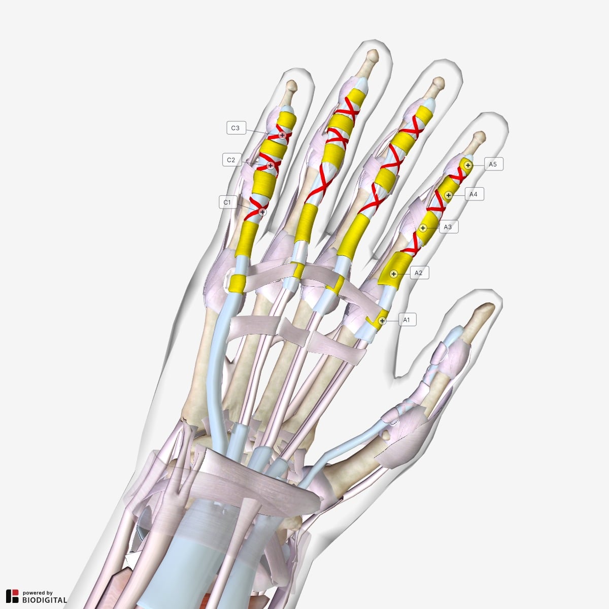
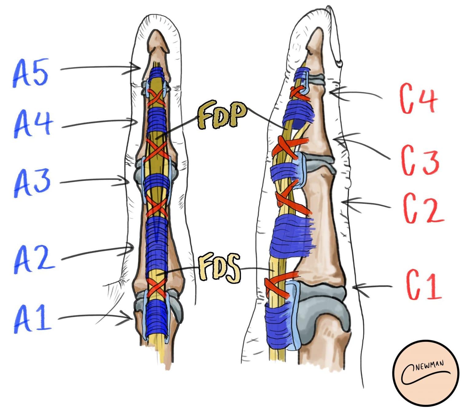
Pathology The annular pulleys are the most functionally important and commonly injured. They comprise a transversely oriented sheath of fibrous tissue that wraps over the flexor superficials and profunda tendons. There are five flexor tendon pulleys in the fingers that are named A1-A5. The thumb only has two pulleys that are described as A1 and A2.

Finger flexor tendon sheath and pulley system Alila Medical Images
The coronal plane is important for evaluating the collateral ligaments and articular cartilage, and the sagittal plane is important for evaluating the flexor and extensor tendons, volar plate, pulleys, and articular cartilage.

a A diagram of the flexor pulley system of the finger with permission... Download Scientific
1 Images Flexor Pulley System-Fingers Annular ligaments A2 and A4 are critical to prevent bowstringing most biomechanically important A1, A3, and A5 overlie the MP, PIP and DIP joints respectively originate from palmar plate A1 pulley most commonly involved in trigger finger Cruciate pulleys

Flexor Pulley System Anatomy, Injury, and Management Journal of Hand Surgery
The flexor pulley system of the hand is a complex structure that co-ordinates flexion of the digits. It consists of: Long flexor tendons - and their associated synovial sheaths. Annular pulleys - 5 associated with each finger, 2 associated with the thumb. Cruciate pulleys - 3 associated with each finger.
Flexor Tendon Repair Surgical Repair TeachMeSurgery
The flexor pulley system of finger of hand consist of localized thickenings of the digital sheath. The index, middle, ring, and small fingers usually contain five annular pulleys (A1 to A5) and three cruciate pulleys (C1 to C3). Wuertzer SD, Pacholke DA. High-resolution 3-T MRI of the fingers: review of anatomy and common tendon and ligament.
